Published on 02/03/2015 by admin
Filed under Internal Medicine
Last modified 22/04/2025
This article have been viewed 1223 times
Case 33
A 60-year-old male with coronary artery disease suffered from an inferior wall myocardial infarction 2 years earlier and underwent staged percutaneous coronary intervention (PCI) of the completely occluded left circumflex and right coronary arteries. He presents now with complaints of chest pain.
Medications: ramipril, isosorbide mononitrate, aspirin, atorvastatin, and clopidogrel.
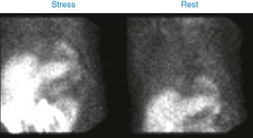
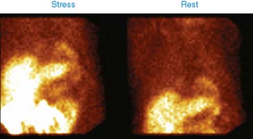
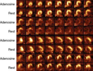
Pharmacologic stress test was performed with 5-minute adenosine infusion. His heart rate changed from 74 to 80 beats/min and blood pressure from 100/60 to 90/60 mm Hg. He had no chest pain or ST changes. He had Q waves in inferior leads, with no change with adenosine infusion. 99mTc-sestamibi was used for stress-rest perfusion imaging.
(Video 1a)
(Video 1b)
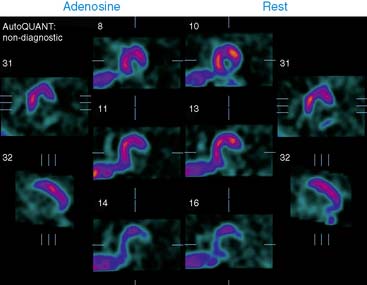
(Fig. 1a)
(Fig. 1b)
(Fig. 1c)
(Video 2)
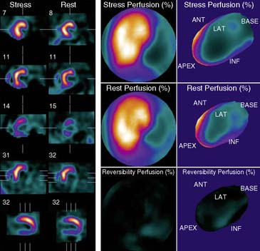
(Fig. 2)
Myocardial perfusion imaging shows a large, dense scar involving the inferior and lateral walls. Left ventricular ejection fraction is 37%. Inferior and lateral walls are akinetic.
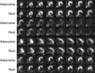
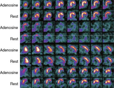
This patient underwent successful PCI of the left circumflex and the right coronary arteries. However, there is a large dense scar involving these vascular territories.
Clinical Nuclear Cardiology State of the Art and Future Directio
WhatsApp us