CASE 33
1. What should be included in the differential diagnosis? (Choose all that apply.)
D. Syphilis
2. What is the most likely diagnosis?
D. Syphilis
3. Which MRI technique could be used to quantify aortic stenosis?
A. Double inversion recovery (black blood)
D. Magnetic resonance angiography (MRA)
4. If the peak velocity across the valve is 3 m/s, what is the pressure gradient?
A. 3 mm Hg
B. 9 mm Hg
C. 12 m Hg
D. 36 mm Hg
ANSWERS
Reference
Walker CM, Reddy GP, Steiner RM. Radiology of the heart Chapter 10. In: Rosendorff C, ed. Essential Cardiology. ed 3 New York: Springer; 2013.
Cross-Reference
Cardiac Imaging: The REQUISITES, ed 3, pp 172–177.
Comment
Etiology
Isolated stenosis of the aortic valve is most commonly secondary to congenital bicuspid valve. Rheumatic heart disease is another important cause of aortic stenosis.
Imaging Features
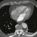
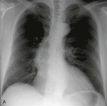
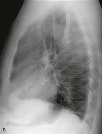
Mild-to-moderate aortic stenosis can cause left ventricular hypertrophy. The left ventricular border may be rounded, or the cardiac apex may be elevated secondary to concentric left ventricular hypertrophy. More severe valvular stenosis can lead to enlargement of the left ventricle and atrium and to hypertrophy. The ascending aortic contour bulges rightward secondary to poststenotic dilation (Fig. A). Calcification of the valve can develop as a result of degeneration and can be seen on CT or, when severe, on chest radiographs (Fig. B).