Published on 02/03/2015 by admin
Filed under Internal Medicine
Last modified 22/04/2025
This article have been viewed 1244 times
Case 31
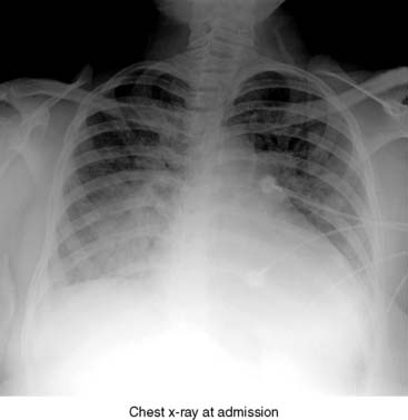
A 19-year-old African American woman who had a normal vaginal delivery 3 weeks earlier developed progressive shortness of breath after delivery. She was found to be in severe heart failure at admission and was diagnosed with postpartum heart failure.
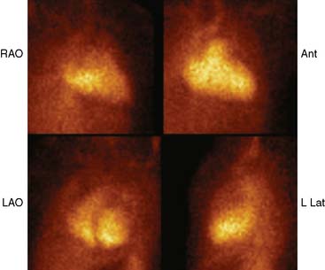
(Fig. 1)
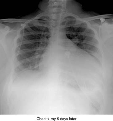
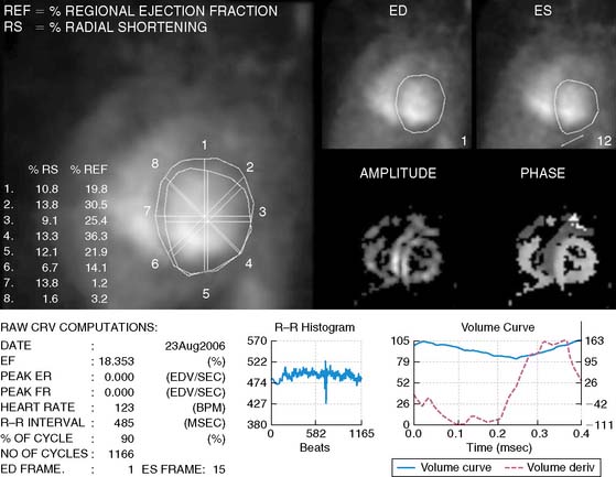
(Fig. 2a)
(Fig. 2b)
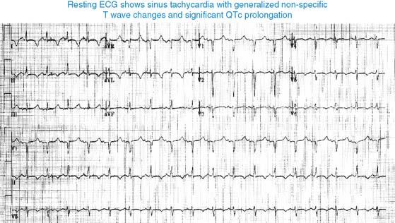
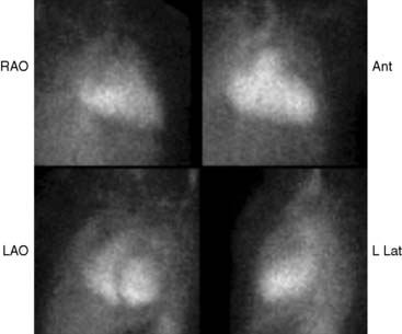
She improved with treatment and is being prepared for discharge. A multiple gated acquisition (MUGA) scan and first-pass imaging were carried out prior to discharge. ECG showed sinus tachycardia with generalized nonspecific T-wave changes and significant QTc prolongation.
(Video 1a)
(Video 1b)
(Fig 2)
On dynamic first imaging, the right atrium and right vent ricle are enlarged and severely hypokinetic, and right ventricular ejection fraction is depressed at 12%. On MUGA, right atrium and right ventricle are markedly enlarged and severely hypokinetic, and left ventricle is massively enlarged and severely hypokinetic. Left ventricular ejection fraction is severely impaired at 18%.
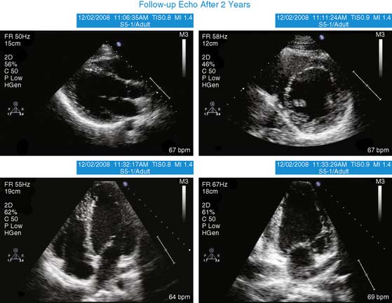
The patient was discharged home and continued to improve after discharge. Echocardiogram 2 years later showed a significant reduction in LV and RV size, and LVEF improved to 40%.
(Video 2)
Peripartum cardiomyopathy generally occurs in the last month of pregnancy or within 5 months of delivery, and its incidence is reported to be 1 in 4000 to 15,000 deliveries in the United States. Clinical course is variable, with 50% to 60% of patients showing significant improvement within the next 6 months; the remaining 40% to 50% show no improvement or deterioration over time.
FURTHER READING
1. Ntusi N.B., Mayosi B.M. Aetiology and risk factors of peripartum cardiomyopathy: A systematic review. Int J Cardiol. 2009;131:168-179.
2. Hilfiker-Kleiner D., Sliwa K., Drexler H. Peripartum ardiomyopathy: Recent insights in its pathophysiology. Trends Cardiovasc Med. 2008;18:173-179.
Clinical Nuclear Cardiology State of the Art and Future Directio
WhatsApp us