CASE 27
1. What should be included in the differential diagnosis? (Choose all that apply.)
B. Ventricular septal defect (VSD)
C. Patent ductus arteriosus (PDA)
D. Partial anomalous pulmonary venous connection (PAPVC)
2. What is the most likely diagnosis?
A. ASD
B. VSD
C. PDA
D. PAPVC
3. What lesion is commonly associated with this anomaly?
A. VSD
B. PDA
C. PAPVC
D. Total anomalous pulmonary venous connection
4. Which anomaly can manifest with cyanosis and shunt vascularity on chest radiographs?
ANSWERS
Reference
Higgins CB. Radiography of congenital heart disease. In: Webb WR, Higgins CB, eds. Thoracic Imaging: Pulmonary and Cardiovascular Radiology. ed 2 Philadelphia: Lippincott Williams & Wilkins; 2010.
Cross-Reference
Cardiac Imaging: The REQUISITES, ed 3, pp 335–338.
Comment
Types of Atrial Septal Defect
There are several types of ASD, including ostium secundum, ostium primum, and sinus venosus. Ostium secundum ASD is the most common type and is the most frequently diagnosed left-to-right shunt in adult patients. Ostium primum ASD is present in atrioventricular septal defect (formerly known as endocardial cushion defect). A sinus venosus defect is commonly associated with PAPVC.
Imaging Findings
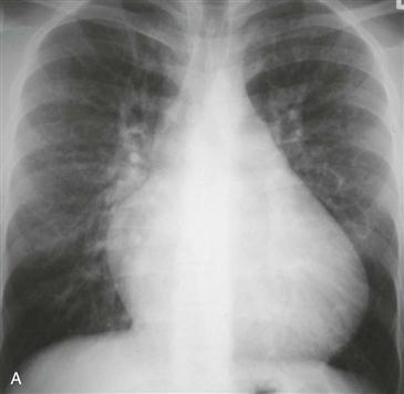
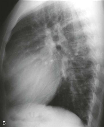
Although the chest radiograph may be normal when the shunt is small, pulmonary vascularity is usually increased (shunt vascularity) (Figs. A and B). Typically, the main pulmonary artery, peripheral pulmonary branches, right atrium, and right ventricle are enlarged. The left atrium is not enlarged; this is an important sign that differentiates ASD from VSD or PDA. PAPVC is another atrial level shunt that can mimic ASD physiologically and can appear similar to ASD on a chest radiograph. Echocardiography can delineate the size and location of the ASD. MRI can be performed if echocardiography does not demonstrate a suspected ASD, and velocity-encoded cine phase contrast MRI can be used to quantify the severity of the shunt.


