Published on 02/03/2015 by admin
Filed under Internal Medicine
Last modified 22/04/2025
This article have been viewed 1085 times
Case 26
A 49-year-old African American woman with hypertension, hyperlipidemia, and smoking suffered from an anterior wall myocardial infarction (MI). She was transferred from a small community hospital and presented to our hospital via MedEvac. She underwent urgent cath, which showed large left anterior descending (LAD) with a large diagonal branch that was completely occluded in the mid-part. The occluded LAD was opened and stented, with no residual lesion. The left circumflex (LCX) was a small nondominant vessel with 50% lesion in the mid-vessel. The right coronary artery (RCA) was also a medium-caliber vessel, with a focal 80% lesion in the mid-segment, followed by 50% narrowing. The LCX and RCA were not angioplastied. She recovered from her MI and was referred for adenosine stress and rest myocardial perfusion imaging 6 weeks later to determine whether she needs revascularization of the RCA lesion.
Medications: lisinopril, Coreg, aldactone, Lipitor, iron, aspirin, and Plavix.
Results of the 5-minute adenosine infusion were changes in heart rate from 68 to 80 beats/min and blood pressure from 151/100 to 135/85 mm Hg. No chest pain was reported.
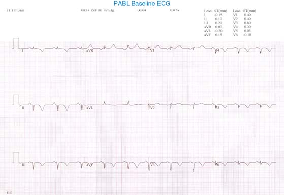
(Fig. 1a)
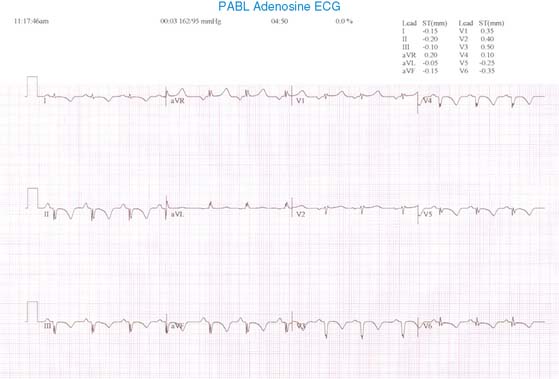
(Fig. 1b)
Normal sinus rhythm with Q waves in precordial leads and in leads II, III, and aVF. No change with adenosine.
(Video 1a)
(Video 1b)
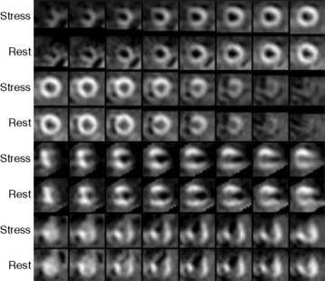
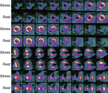
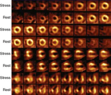
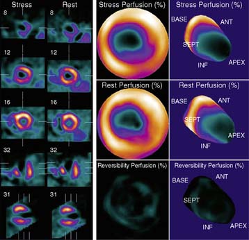
(Fig. 2a)
(Fig. 2b)
(Fig. 2c)
(Fig. 3a)
(Fig. 3b)
(Video 2a)
(Video 2b)
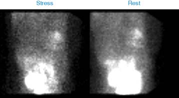
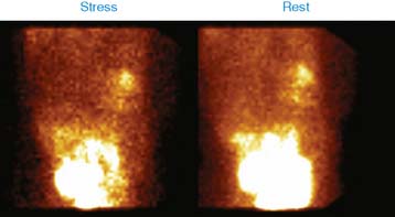
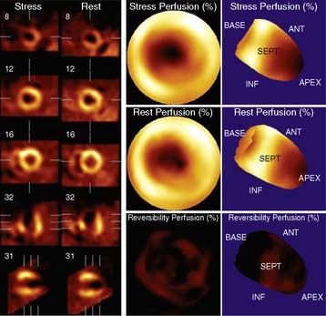
There is a large dense area of perfusion abnormality involving the distal part of the anterior wall, septum, apex, and contiguous inferior wall. The anterior wall and septum are hypokinetic, and the apex is dyskinetic. Left ventricular ejection fraction (LVEF) is impaired at 39%.
Months later, the patient presented with recurrence of chest pain and was ruled in for a small non-ST-segment elevation MI. Repeat coronary angiography showed 40% in-stent narrowing of the LAD, and RCA had 90% mid-narrowing and 100% distal narrowing. Both lesions were successfully angioplastied and stented.
Her LVEF was impaired at 25%. She also received an implantable cardioverter defibrillator (ICD) for primary prevention of sudden cardiac death.
Clinical Nuclear Cardiology State of the Art and Future Directio
WhatsApp us