CASE 26
1. What cardiac chambers are enlarged? (Choose all that apply.)
A. Left atrium
C. Right atrium
2. What is the most likely diagnosis?
3. What is the most common cause of papillary muscle rupture?
A. Trauma
D. Iatrogenic
4. What is the major valvular lesion in acute rheumatic fever?
ANSWERS
Reference
Walker CM, Reddy GP, Steiner RM. Radiology of the heart Chapter 10. In: Rosendorff C, ed. Essential Cardiology. ed 3 New York: Springer; 2013.
Cross-Reference
Cardiac Imaging: The REQUISITES, ed 3, pp 190–194.
Comment
Pathology and Etiology
Mitral regurgitation is caused by a malfunction of any part of the mitral apparatus, including the valve leaflets, chordae, papillary muscles, mitral annulus, and adjacent left ventricular wall. Causes of mitral regurgitation include ischemic cardiomyopathy, myocardial infarction, papillary muscle rupture, rheumatic heart disease, endocarditis, and trauma.
Imaging Findings and Diagnostic Criteria
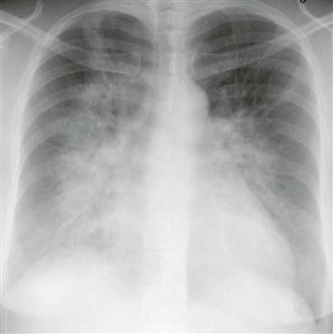
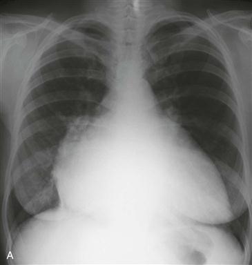
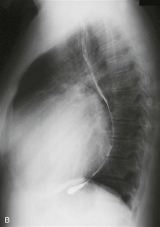
Chest radiographic findings of mitral regurgitation vary depending on the chronicity and severity of disease. Acute severe mitral regurgitation results in pulmonary venous hypertension and alveolar edema without significant cardiac enlargement. After several days, the heart dilates, and interstitial edema persists. After weeks to months, the left atrium and ventricle are enlarged (Figs. A and B), and the pulmonary pattern is variable.
Echocardiography is commonly used to grade the severity of mitral regurgitation. MRI can be used to quantify the regurgitant fraction.