CASE 25

History: A 35-year-old man presents with 3 weeks of upper abdominal pain.
1. Which of the following should be included in the differential diagnosis of the dominant imaging findings? (Choose all that apply.)
2. What is the most likely lesion to cause large masses involving the gut without bowel obstruction?
3. Which of the following correctly describes aneurysmal dilation of the small bowel?
B. Dilation of the superior mesenteric artery
C. Dilation of the small bowel proximal to a segment of narrowing
D. A diverticulum greater than 10 cm in diameter
4. What small bowel lesion results in elevated levels of 5-hydroxyindoleacetic acid (5-HIAA) in the urine?
ANSWERS
CASE 25
Small Bowel Lymphoma
1. A, B, C, and E
2. B
3. A
4. C
References
Dahnert W. Radiology Review Manual. 6th ed. Philadelphia: Lippincott Williams & Wilkins; 2007. pp 851-852
Cross-Reference
Gastrointestinal Imaging: THE REQUISITES, 3rd ed, p 133.
Comment
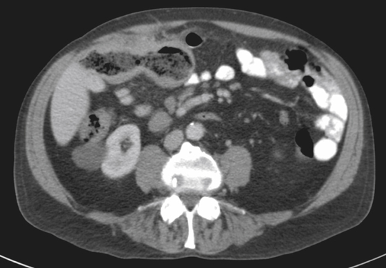
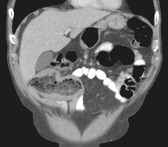
Lymphoma is a common malignancy of the small bowel. It can grow to large masses, which, quite often, do not obstruct the bowel. This phenomenon is due to a particular characteristic of lymphoma called aneurysmal dilation, in which the gut lumen is channeled through the lesion, leaving a false channel surrounded by tumor but without obstruction (see figures). This condition may be rarely seen in other small cell lesions of the gut, such as melanoma, but it most commonly indicates lymphoma. It does not seem to occur in Hodgkin’s disease, and the mechanism is not understood. As a result, patients can present with large abdominal masses, possibly with constitutional symptoms, but no complaints related to bowel obstruction. CT findings include large masses involving the bowel, most often with large foamy lymphomatous nodes in the peritoneum, in the retroperitoneum, and at the base throughout the mesentery (see figures).


