[level-membership-for-gastroenterology-and-hepatology-category]CASE 24
History: A 29-year-old man presents with dizziness and dyspepsia.
1. Which of the following should be included in the differential diagnosis of the dominant imaging finding on figures A and B? (Choose all that apply.)
E. Postinflammatory pseudocysts
2. Several abdominal lesions have been reported to be associated with von Hippel–Lindau disease (VHL). Which of the following is not a recognized association?
D. Pancreatic islet cell tumor
3. Which of the following studies would be most appropriate in the work-up of this patient?
4. Regarding VHL disease, which of the following statements is true?
A. Cerebellar hemangioblastomas are usually multiple and aggressive.
B. Pheochromocytoma in VHL disease occurs in older patients and is usually multiple.
C. The renal malignancies associated with VHL disease are clear cell tumors.
D. The scrotal lesions associated with VHL disease are testicular cystadenomas.
ANSWERS
CASE 24
von Hippel–Lindau Disease
1. C, D, and E
2. C
3. D
4. C
Reference
Choyke PL, Glenn GM, Walther MM, et al: von Hippel-Lindau disease: genetic, clinical and imaging features. Radiology. 1995;194:629–642.
Cross-Reference
Gastrointestinal Imaging: THE REQUISITES, 3rd ed, p 168.
Comment
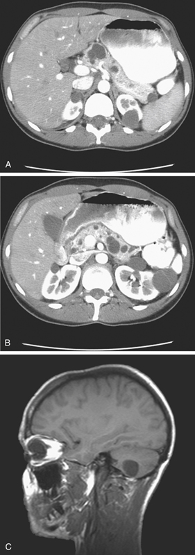
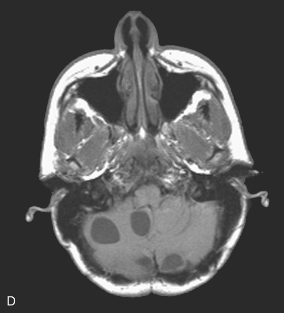
The images in this case show multiple pancreatic cysts of varying size (see figures). The CT images are from a patient with von Hippel–Lindau (VHL) disease. VHL disease is an autosomal dominant inherited disease of capillary angiomatous hamartomas with CNS involvement of the brain and retina. In the brain, the lesion is usually a hemangioblastoma of the posterior fossa (see figures). Visceral abdominal tumors also seen in this condition include renal cell carcinoma, pheochromocytoma, and increased incidence of cystic malignancy of the pancreas.
Multiple pancreatic cysts are seen in almost three quarters of patients with VHL disease and are highly suggestive of the diagnosis. Solid islet cell tumors may be seen in VHL disease. Most of the cases are discovered in the third and fourth decades of life. If malignancy is present at the time of diagnosis, the prognosis is usually poor. Thus, suspicion should be aroused when any patient undergoing a CT of the abdomen demonstrates multiple pancreatic cysts, and the abdomen should be carefully searched for such neoplasms as pheochromocytomas and renal cell cancers.
[/level-membership-for-gastroenterology-and-hepatology-category][not-level-membership-for-gastroenterology-and-hepatology-category]CASE 24
History: A 29-year-old man presents with dizziness and dyspepsia.
1. Which of the following should be included in the differential diagnosis of the dominant imaging finding on figures A and B? (Choose all that apply.)
E. Postinflammatory pseudocysts
2. Several abdominal lesions have been reported to be associated with von Hippel–Lindau disease (VHL). Which of the following is not a recognized association?
D. Pancreatic islet cell tumor
[/not-level-membership-for-gastroenterology-and-hepatology-category]
