CASE 23
History: An 82-year-old woman presents with heartburn.
1. Which of the following should be included in the differential diagnosis of the dominant imaging finding on figure A? (Choose all that apply.)
2. Review the figures. What is the most likely diagnosis?
A. Aberrant left subclavian artery
B. Aberrant right subclavian artery
C. Anomalous superior vena cava
3. How does a patient with an aberrant right subclavian artery usually present?
4. Which of the following is not an extrinsic impression normally seen during a barium swallow?
ANSWERS
CASE 23
Aberrant Right Subclavian Artery
1. A, B, D, and E
2. B
3. A
4. C
References
Berdon WE. Rings, slings, and other things: vascular compression of the infant trachea updated from the midcentury to the millennium—the legacy of Robert E. Gross, MD, and Edward B.D. Neuhauser, MD. Radiology. 2000;216:624–632.
Freed K, Low VHS. The aberrant subclavian artery. Am J Roentgenol. 1997;168(2):481–484.
Cross-Reference
Gastrointestinal Imaging: THE REQUISITES, 3rd ed, p 12-14.
Comment
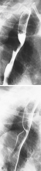
The normal extrinsic impressions one expects to see on the esophagus are the aortic arch, the left main bronchus, and the left atrium of the heart. Congenital malplaced right subclavian artery originating from the distal aspect of the aortic arch (instead of its usual origin off the right common carotid artery) is seen in slightly less than 1% of the population. In crossing back from left to right across the mediastinum to reach the right limb, the aberrant right subclavian artery passes behind the esophagus. The posterior-crossing artery shown here impressed the posterior wall of the upper esophagus and may produce dysphagia.
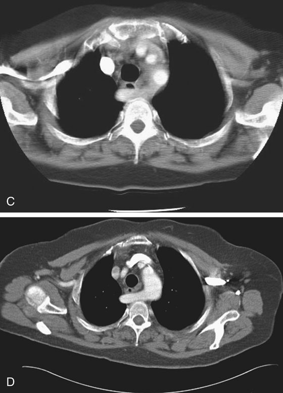
A barium esophagogram is the simplest and least expensive way of making the diagnosis (see figures). However, if necessary, multidetector computed tomography (MDCT) can demonstrate the anomaly, as in the images presented in this case (see figures). Here one can see the aberrant subclavian artery cross to the right of the mediastinum behind the esophagus. The term dysphagia lusoria historically applied to the impression of a greatly enlarged left atrium of rheumatic heart disease. However, today it is generally used to describe dysphagia caused by any impression by any vascular structure.