Case 23

Published on 02/03/2015 by admin

Filed under Internal Medicine

Last modified 22/04/2025

Print this page

rate 1 star rate 2 star rate 3 star rate 4 star rate 5 star
Your rating: none, Average: 0 (0 votes)

This article have been viewed 1353 times

Case 23

A 61-year-old male with hypertension, hyperlipidemia, and known coronary artery disease was admitted to the hospital with worsening congestive heart failure. He suffered from a myocardial infarction in the past, for which he underwent percutaneous transluminal coronary angioplasty (PTCA). He was referred for pharmacologic stress perfusion imaging prior to consideration for a biventricular pacing ICD.

Medications: aspirin, furosemide, lisinopril, carvedilol, atorvastatin, spironolactone, and digoxin.

He underwent pharmacologic stress using 5-minute adenosine infusion protocol. His heart rate changed from 63 to 90 beats/min and blood pressure from 115/71 to 133/76 mm Hg. There was no chest pain. Baseline and peak stress ECGs are shown next. What is your interpretation?

There is normal sinus rhythm with left bundle branch block and markedly prolonged QRS. QRS measures more than 180 msec. There is no change with adenosine infusion.

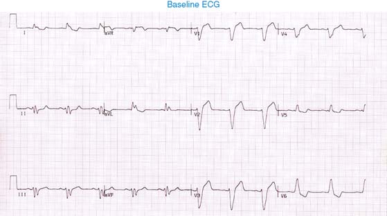
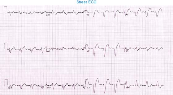
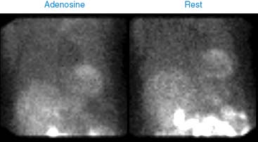
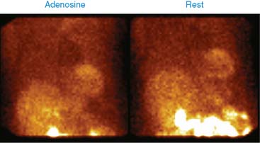
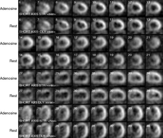
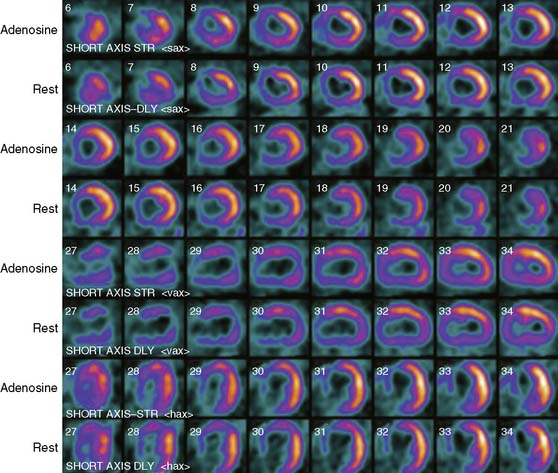
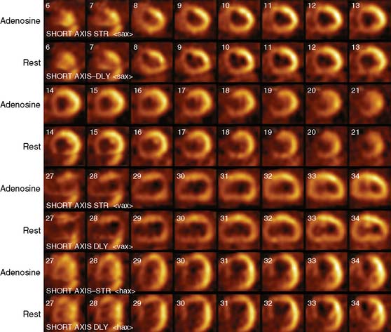
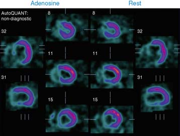
His 99mTc-sestamibi stress and rest images are shown next. What is your interpretation?

The LV is massively enlarged. There is a large, dense area of perfusion abnormality involving the septum and part of the inferior wall and anterior wall. There is no change on rest images. On gated SPECT images, the septum is dyskinetic, and the remaining LV is severely hypokinetic. LVEF is severely reduced at 23%.

He received BiV-ICD for ischemic cardiomyopathy. During subsequent outpatient follow-ups, his heart failure has remained stable.