CASE 23
1. What tumors can invade the inferior vena cava (IVC)? (Choose all that apply.)
B. Hepatocellular carcinoma (HCC)
2. What is the cardiac finding?
3. What is the source of tumor thrombus?
B. HCC
D. Sarcoma
4. Which of the following is characteristic of a malignant cardiac mass?
B. Infiltrative
ANSWERS
CASE 23
Reference
Dedeilias P, Nenekidis I, Koukis I, et al. Acute heart failure caused by a giant hepatocellular metastatic tumor of the right atrium. J Cardiothorac Surg. 2011;6:102.
Cross-Reference
Cardiac Imaging: The REQUISITES, ed 3, pp 277–278.
Comment
Imaging
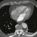
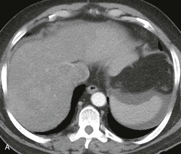
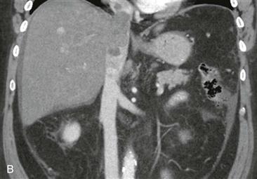
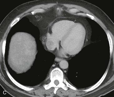
Multiple images from a contrast-enhanced CT scan of the abdomen show a heterogeneously enhancing filling defect expanding the middle hepatic vein and IVC (Figs. A and B). The filling defect extends superiorly into the right atrium (Fig. C). These imaging findings are most consistent with tumor thrombus from HCC. Patients with HCC uncommonly present with metastatic disease (approximately 5%).
Overview
Invasion of the IVC and right atrium by tumor is uncommon. The three most common tumors to invade the IVC and right atrium are HCC, renal cell carcinoma, and adrenocortical carcinoma. Cardiac metastases secondary to HCC occur in 3% of all patients. Prompt diagnosis is important because tumor within the right atrium can cause congestive heart failure and sudden cardiac death. Surgical resection is palliative but rarely curative.