CASE 22

History: A 62-year-old woman presents with dyspnea and abdominal distention; she has a history of ovarian carcinoma.
1. Which of the following should be included in the differential diagnosis of the dominant imaging finding on figure A? (Choose all that apply.)
A. Acquired (post-traumatic) pseudocyst
2. What is the most common cause for benign splenic cysts?
3. Which of the following is true about metastatic disease involving the spleen?
A. Lymphomas are the most common splenic malignancy.
B. Metastatic ovarian cancer is cystic owing to central necrosis.
C. The most common origin of splenic metastases is melanoma.
D. 25% to 50% of patients with terminal disease owing to metastatic malignancy have splenic lesions.
4. What is the most common benign neoplasm of the spleen?
ANSWERS
CASE 22
Metastatic Disease to the Spleen
1. A, C, D, and E
2. D
3. A
4. C
References
Kamaya A, Weinstein S, Desser TS. Multiple lesions of the spleen. Semin Ultrasound CT MRI. 2006;27:389–403.
Warshauer DM, Hall HL. Solitary splenic lesions. Semin Ultrasound CT MRI. 2006;27:370–388.
Cross-Reference
Gastrointestinal Imaging: THE REQUISITES, 3rd ed, p 215.
Comment
The most common cystic lesions that affect the spleen are the acquired lesions and epidermoid cysts. Acquired cysts are thought to be mostly traumatic in origin (the small splenic laceration that heals, leaving a hematoma, seroma, and finally a cystic fluid collection that can remain indefinitely). These cysts are without a well-defined lining and calcify over the years. They account for about 80% of benign cystic lesions of the spleen and are best left alone. Epidermoid cysts have a well-defined epithelial lining and are probably congenital in origin. These cysts are also often an incidental finding on CT examinations. Occasionally a pseudocyst in the pancreatic tail can be confused with a splenic cyst. However, with multidetector computed tomography (MDCT) and multiplanar evaluation of the abdomen, these can usually be sorted out.
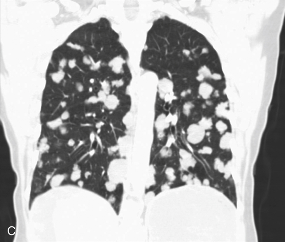
Primary neoplasms of the spleen are rare, with hemangioma being the most common. Metastatic disease involving the spleen is also very uncommon. However, as seen in the images, it should not be neglected in the differential diagnosis for splenic defects in patients with known primary neoplasms (see figures). In this case, the primary cancer is ovarian carcinoma, and there has been widespread dissemination of tumor throughout the peritoneal cavity with ascites (see figures) and invasion of the spleen as well as involvement of the thoracic cavity (see figures).