CASE 22
1. What are life-threatening complications of this disease? (Choose all that apply.)
A. Tamponade
B. Stroke
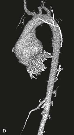
2. What is the term used to describe the appearance of this ascending aorta?
3. Which of the following conditions is most commonly associated with annuloaortic ectasia?
4. Which complication of annuloaortic ectasia has occurred in this patient?
C. Tamponade
ANSWERS
Reference
Ha HI, Seo JB, Lee SH, et al. Imaging of Marfan syndrome: multisystemic manifestations. Radiographics. 2007;27(4):989–1004.
Cross-Reference
Cardiac Imaging: The REQUISITES, ed 3, pp 378–391.
Comment
Imaging
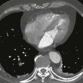
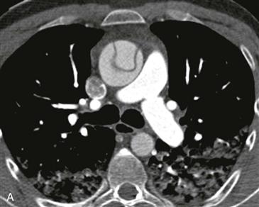
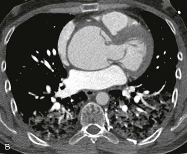
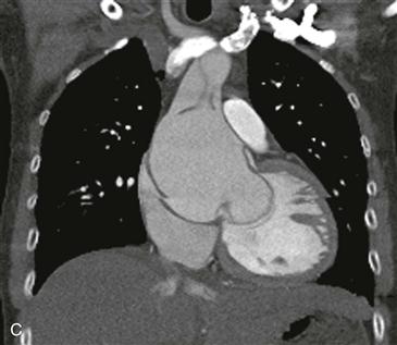
Axial and coronal images from prospectively gated CT angiography of the thoracic aorta show a Stanford type A aortic dissection (Figs. A–C). The intimal flap is prolapsed through the aortic valve causing aortic regurgitation (Figs. B and C). The ascending aorta is shaped like a pear or tulip bulb (Figs. C and D), which is consistent with annuloaortic ectasia. Annuloaortic ectasia is most commonly seen in Marfan syndrome but can occur in other connective tissue diseases such as Ehlers-Danlos syndrome.
Complications
Annuloaortic ectasia occurs secondary to cystic medial necrosis, which may be idiopathic or associated with Marfan syndrome or Ehlers-Danlos syndrome. Three main complications of this condition include aortic rupture, aortic regurgitation, and aortic dissection. Early operative repair of a thoracic aortic aneurysm is performed when the diameter reaches 5 cm in patients with Marfan syndrome because of the high risk of rupture.