CASE 21
History: A 77-year-old woman had undergone transvaginal uterine polypectomy 2 days ago. She complains of nausea, syncope, and hematochezia.
1. Which of the following should be included in the differential diagnosis of the imaging findings shown? (Choose all that apply.)
2. What is the most likely diagnosis in this case?
3. All of the following CT findings may be associated with ischemic colitis except:
4. Which of the following describes the prognosis of ischemic colitis?
A. Most patients improve and fully recover.
B. Most patients develop progressive colonic gangrene.
C. Most patients develop a stricture.
D. Patients are at high risk for developing colon carcinoma.
ANSWERS
CASE 21
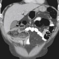
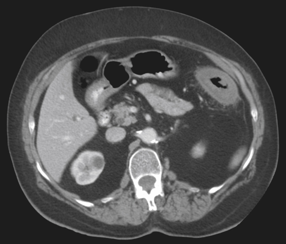
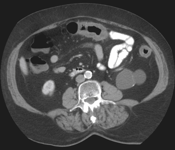
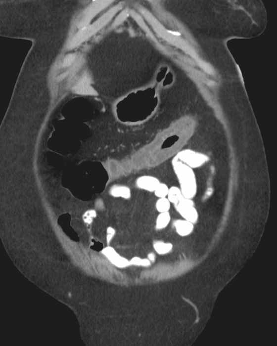
Ischemic Colitis of the Transverse Colon
1. A, B, D, and E
2. D
3. C
4. A
References
Rha SE, Ha HK, Lee SH, et al: CT and MR imaging findings of bowel ischemia from various primary causes. Radiographics. 2000;20:29–42.
Cross-Reference
Gastrointestinal Imaging: THE REQUISITES, 3rd ed, p 298.
Comment
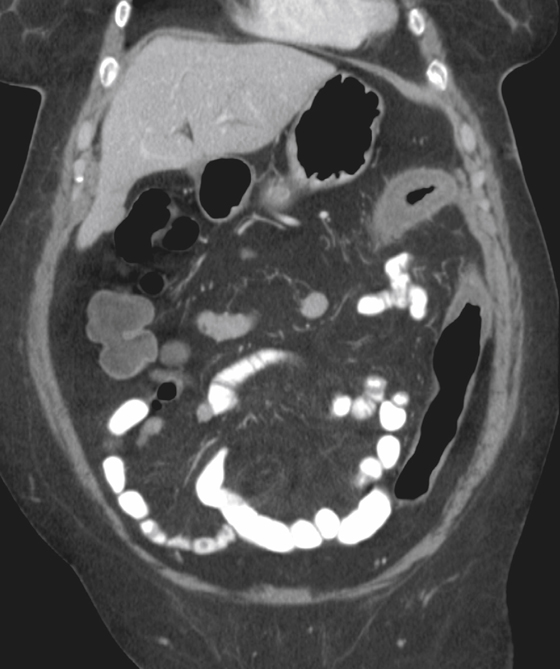
Ischemic bowel disease is a common clinical problem, especially in the elderly. Most often it is produced by low flow to the intestines, which occurs in patients who are in hypotensive states, those who are experiencing cardiac failure, and patients who have just had surgery, and in patients with other conditions. Arterial obstruction is less common but can affect elderly patients with atherosclerotic disease that obstructs mesenteric vessels or those with embolic disease who have cardiac abnormalities. Also, a small percentage of patients have ischemia caused by venous obstruction.
Arteriography is being used less commonly with the advent of high-quality multidetector computed tomography (MDCT) in patients with ischemia. Angiography often does not demonstrate obstructing vessels. CT is probably the best modality for defining the involved segments and identifying underlying pathologic conditions or complications (see figures).
Ischemic disease of the colon is often segmental and rarely involves the entire colon (see figures). The different regions of the colon have separate blood supplies, although they are interconnected to some extent. The watershed regions are defined as the areas of transition between the superior and inferior mesenteric blood supplies. This term often applies to the splenic flexure (see figures), although sometimes it can include parts of the rectosigmoid junction as well. The splenic flexure is thought by some to be the most susceptible area for ischemia in patients with low flow-states because it is the most distal point of arterial flow to the colon. On the other hand, some think that being supplied by two arterial systems confers some protection.
Interestingly, most disease seems to occur in the descending and sigmoid regions. The right colon also is commonly involved because it is prone to ischemia resulting from pathologic distention. The right colon is the most distended segment of colon under most conditions.
Radiologically, ischemia first can be identified as thickening and edema of the bowel wall. Nodularity of the bowel wall could be the result of either multiple areas of focal edema or hemorrhage (thumbprinting). The mucosa may become shaggy and begin to resemble inflammatory bowel disease. As the ischemic area heals, the colon may become fibrotic, with loss of haustral pattern and even with formation of pseudodiverticula. This fibrosis can be multifocal. However, in at least 50% of cases healing is complete without stricture.