Case 21

Published on 02/03/2015 by admin

Filed under Internal Medicine

Last modified 22/04/2025

Print this page
rate 1 star rate 2 star rate 3 star rate 4 star rate 5 star
Your rating: none, Average: 0 (0 votes)

This article have been viewed 1319 times

Case 21

A 45-year-old Caucasian male who is a truck driver and hauls sand and stones started experiencing palpitations and shortness of breath 2 months previously, which progressively worsened to four-pillow orthopnea. He is a heavy drinker (6 to 12 beers per day) and chronic smoker. He was admitted to an outside hospital in frank pulmonary edema, where he underwent catheterization after diuresis.

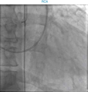
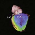
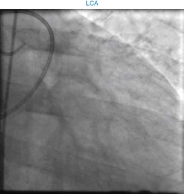
He was found to have complete occlusion of the LCX, severe disease of the D1 and D2, normal RCA, and aneurysm of the lateral wall. He was referred to our hospital for further evaluation and surgical treatment.

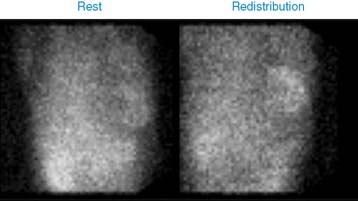
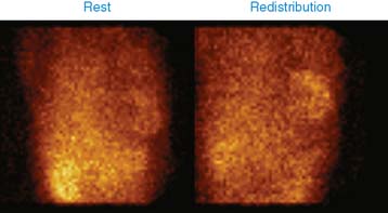
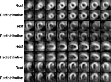
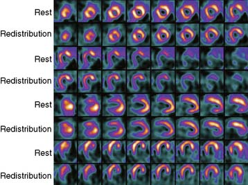
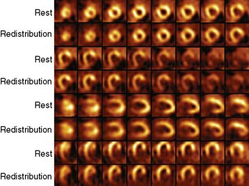
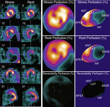
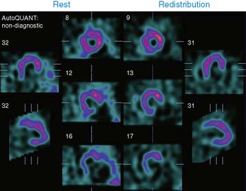
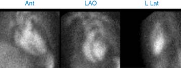
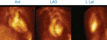
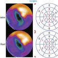
During the course of subsequent evaluation, a viability study was requested. An injection of 3.2 mCi of thallium-201 (Tl-201) was given, and gated SPECT imaging was done 15 minutes and 4 hours later. A large dense scar involving the inferior and lateral wall was noted, with no evidence of viability. Tracer uptake was normal in the remaining left ventricle, indicating the presence of normal viability in the remaining ventricle. LVEF was 35%. There is a suspicion of a large pseudoaneurysm arising from the lateral wall on this study, because of the presence of a large dense photopenic area adjoining the lateral wall. He underwent MUGA scan.

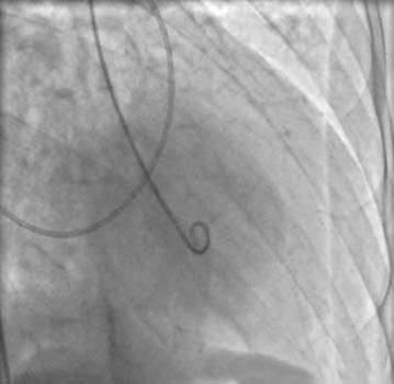
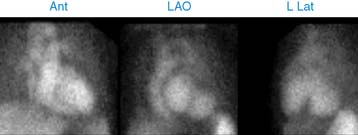
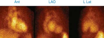
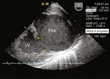
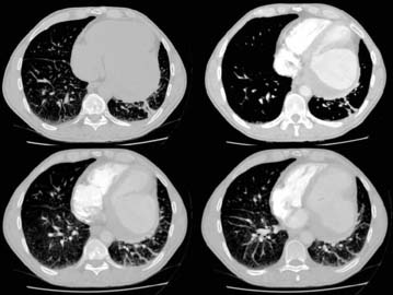
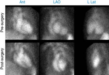
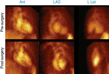
The right atrium and right ventricle were normal, but MUGA showed a massive pseudoaneurysm arising from the lateral and inferobasal walls. The remaining LV contracts normally and vigorously. The EF of the LV after exclusion of the pseudoaneurysm is 36%. TEE and CT scan with and without contrast were performed to further evaluate the pseudoaneurysm prior to surgery. Contrast CT showed large mural thrombi within the pseudoaneurysm.

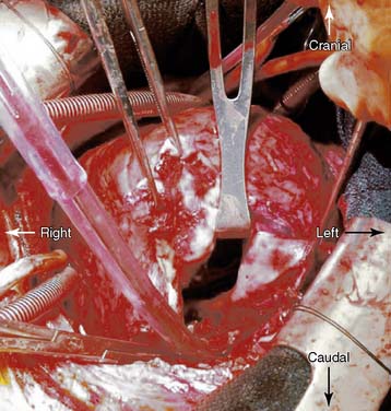
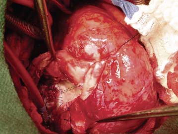
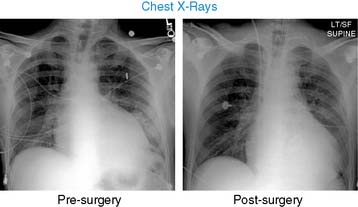
He underwent surgery for aneurysmectomy and coronary bypass grafts. A large lateral wall pseudoaneurysm was excised. The lateral wall required a patch, MV was replaced, and OM1, OM2, D1, and D2 bypassed. He had an uneventful postoperative recovery.

Left ventricular pseudoaneurysm is a rare complication following Ac myocardial infarction. This follows rupture of infarcted myocardium, which is contained by adherent pericardium. This is more common in inferior, lateral, and apical infarctions, whereas true aneurysms are more common with anterior infarcts. Untreated pseudoaneurysms carry a high risk of spontaneous rupture. MUGA is a highly reliable technique for the detection of pseudoaneurysm of the LV.