CASE 21

1. What are potential causes of this entity? (Choose all that apply.)
A. Idiopathic
B. Alcohol
D. Infection
E. Duchenne muscular dystrophy
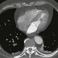
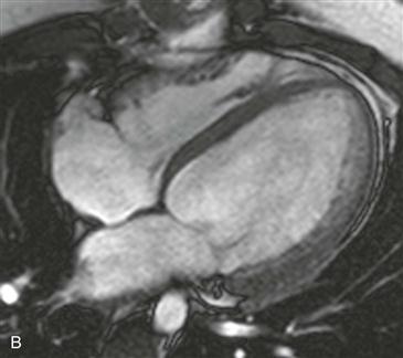
2. What is the finding on the precontrast bright blood sequences? (Fig. A was obtained at end-diastole, and Fig. B was obtained at end-systole.)
A. Dilated cardiac chambers and low ejection fraction
C. Concentric myocardial hypertrophy
D. Spin dephasing flow void artifact
3. What is the most common cause of congestive heart failure?
D. Myocarditis
4. What is the likely cause of this patient’s congestive heart failure?
A. Amyloidosis
ANSWERS
Reference
Sparrow PJ, Merchant N, Provost YL, et al. CT and MR imaging findings in patients with acquired heart disease at risk for sudden cardiac death. Radiographics. 2009;29(3):805–823.
Cross-Reference
Cardiac Imaging: The REQUISITES, ed 3, pp 87–88, 284–286.
Comment
Overview
Nonischemic dilated cardiomyopathy is the third most common cause of heart failure after ischemic and valvular heart disease. It is caused by various insults, including alcohol, medications, infections, and various neuromuscular syndromes. Frequently, it is familial or idiopathic. Histologically, there is loss of myocytes with progressive deposition of interstitial fibrosis leading to impaired ventricular contraction.
Imaging
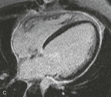
Cine steady-state free precession (SSFP) four-chamber view images show dilation of all four cardiac chambers (Figs. A and B). There is a severely reduced ejection fraction, which was measured at 14%. Late gadolinium enhancement is noted in the midportion of the interventricular septum (Fig. C).
Presentation
Patients usually present with congestive heart failure, arrhythmias, or cardiac thromboembolism secondary to ventricular hypocontractility. Imaging findings include ventricular dilation and impaired myocardial contractility with reduction in ejection fraction. Midmyocardial late gadolinium enhancement is seen in 12% to 35% of patients (Fig. C).
Prognosis and Treatment
Patient mortality is due to heart failure or sudden cardiac death from ventricular arrhythmia. Specific imaging risk factors that convey an increased risk for sudden cardiac death include a reduced left ventricular ejection fraction (<35%) and late gadolinium myocardial enhancement (affecting 25% to 75% of the myocardial wall). Treatment is symptomatic management of heart failure. Heart transplantation is the definitive therapy.