•
Defect in hepatocellular secretion of conjugated bilirubin
•
Autosomal recessive
•
Mutations in
ABCC2 (
CMOAT /
MRP2) gene, which codes for ATP-dependent organic anion transport localized to canalicular membrane
Results in impaired biliary canalicular transport of organic anions including conjugated bilirubin
Impaired glutathione excretion reduces bile salt-independent bile flow
•
Incidence
Rare
•
Age
Develop jaundice in teenage years
•
Sex
M = F
•
Ethnicity
Prevalence highest among Moroccan and Iranian Jews (1:1,300)
•
Most patients asymptomatic
•
Can present as chronic or intermittent jaundice or with mild right upper quadrant abdominal pain
[level-membership-for-pathology-category]
•
Serum bile acids are not increased, so pruritus is absent
•
Urine may be darker than normal
•
Some neonates present with cholestasis
•
Jaundice can be precipitated by pregnancy or by drugs that decrease hepatic excretion of organic anions (e.g., oral contraceptives)
•
Measurement of urine coproporphyrin isomers shows shift from isomer III to isomer I
•
Conjugated hyperbilirubinemia
•
Normal alkaline phosphatase and γ-glutamyl transpeptidase
•
Grossly, liver is darkly pigmented and can appear green, slate blue, dark gray, or black
•
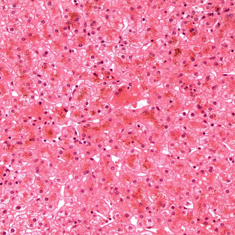
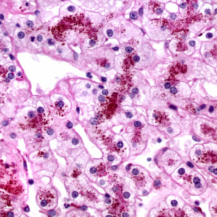
Coarse granular pigment in centrizonal hepatocytes
•
Pigment was earlier thought to be form of melanin or lipofuscin
•
Likely composed of polymers of epinephrine metabolites
•
No other histologic changes
•
PAS with diastase digestion
Accentuates cytoplasmic pigment
•
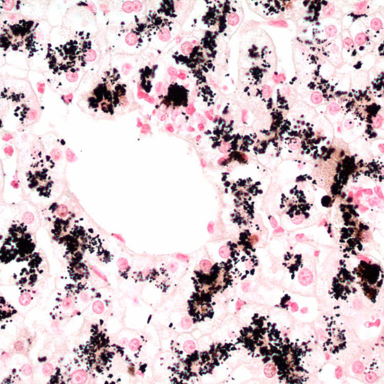
Fontana-Masson
Silver stain that stains cytoplasmic granules black
•
MRP2: Absence of staining of canalicular membrane
Available through referral centers
Helpful in young children whose livers have not accumulated pigment
•
Membrane-bound, electron-dense lysosomal granules within cytoplasm of hepatocytes
•
Can also show grossly pigmented liver but has distinct clinical and histologic features
•
Pigment in centrizonal hepatocytes is not as coarse
•
Unconjugated hyperbilirubinemia
•
Inspissated bile in canaliculi
•
Swelling (feathery degeneration) of hepatocytes in cholestatic area
•
Prussian blue (+) pigment in periportal hepatocytes
•
Coarse pigment in centrizonal hepatocytes in patient with isolated conjugated hyperbilirubinemia
•
Pigment may disappear during episode of hepatitis and reaccumulate after recovery
1.Nisa, AU, et al. Dubin-Johnson syndrome. J Coll Physicians Surg Pak . 2008; 18(3):188–189.
2.Mor-Cohen, R, et al. Age estimates of ancestral mutations causing factor VII deficiency and Dubin-Johnson syndrome in Iranian and Moroccan Jews are consistent with ancient Jewish migrations. Blood Coagul Fibrinolysis . 2007; 18(2):139–144.
3.Jedlitschky, G, et al. Structure and function of the MRP2 (ABCC2) protein and its role in drug disposition. Expert Opin Drug Metab Toxicol . 2006; 2(3):351–366.
4.Lee, JH, et al. Neonatal Dubin-Johnson syndrome: long-term follow-up and MRP2 mutations study. Pediatr Res . 2006; 59(4 Pt 1):584–589.
5.Rastogi, A, et al. Dubin-Johnson syndrome–a clinicopathologic study of twenty cases. Indian J Pathol Microbiol . 2006; 49(4):500–504.
6.Sobaniec-Lotowska, ME, et al. Ultrastructure of Kupffer cells and hepatocytes in the Dubin-Johnson syndrome: a case report. World J Gastroenterol . 2006; 12(6):987–989.
[/level-membership-for-pathology-category][not-level-membership-for-pathology-category]
[/not-level-membership-for-pathology-category]
Diagnostic Pathology Hepatobiliary and Pancreas
 corresponding to the pigment within centrizonal hepatocytes.