[level-membership-for-radiology-category] a.k.a. von Meyenburg complex


 , representing biliary hamartomas. This patient also had evidence of congenital hepatic fibrosis on imaging and liver biopsy, both part of the congenital hepatic and renal fibropolycystic disease spectrum.
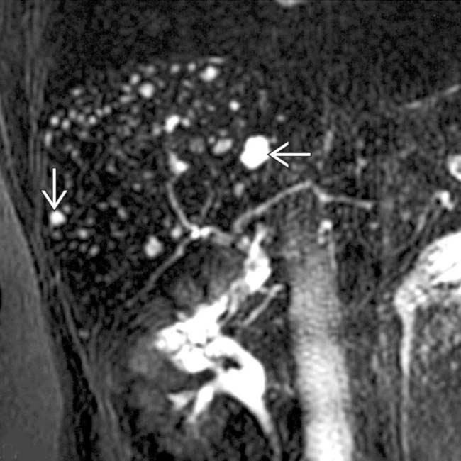
 that do not communicate with the (normal) biliary tree. This feature helps to distinguish biliary hamartomas from Caroli disease.
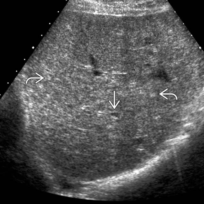
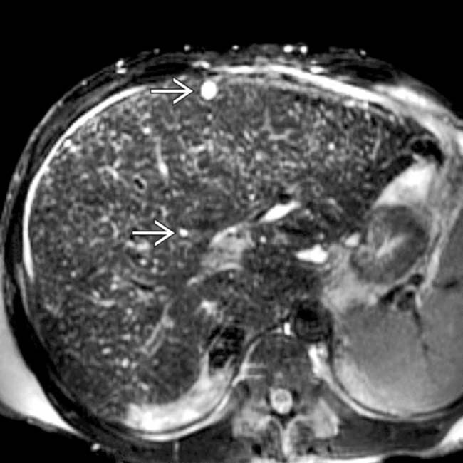
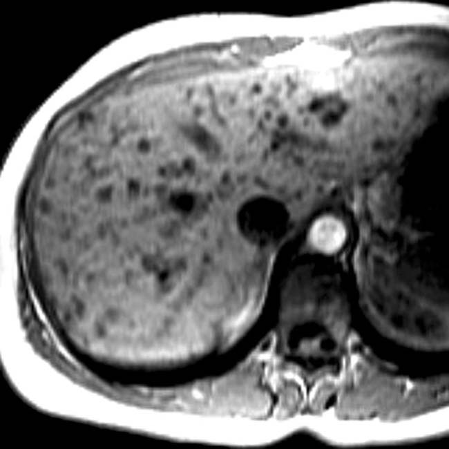
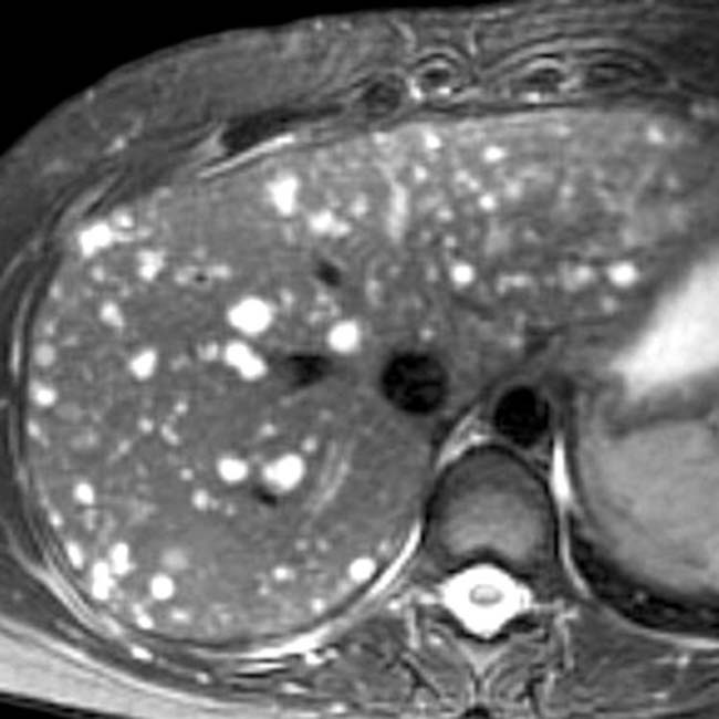
 throughout the liver and 1 of ∼ 10 cyst-like lesions
  , though even these have small foci of echogenicity within the wall. MR on this patient showed many more cystic-appearing biliary hamartomas.
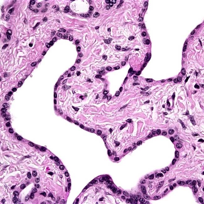


PATHOLOGY
General Features


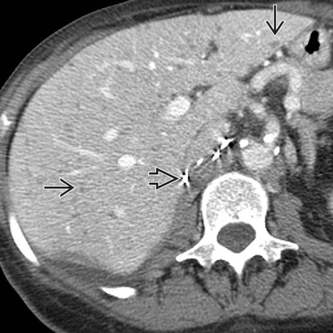
 throughout the liver. Incidental note is made of surgical clips
  from the prior nephrectomy. These biliary hamartomas should not be mistaken for metastases.
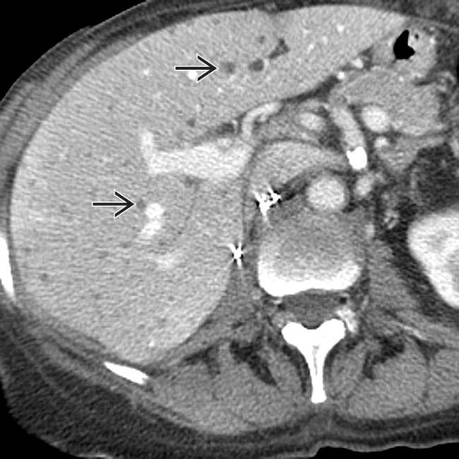
 that almost certainly represent biliary hamartomas. These have remained stable for years, and the patient is not immunosuppressed.
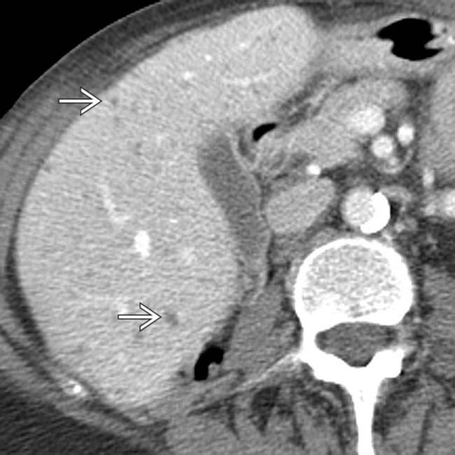
 .
.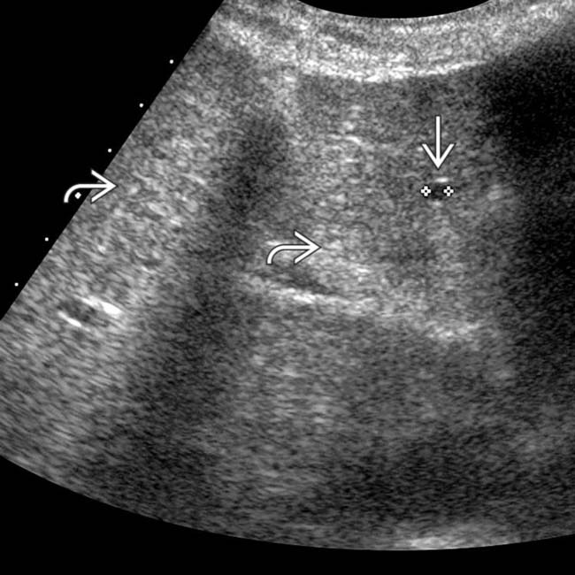
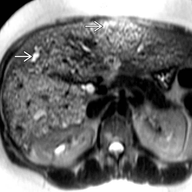
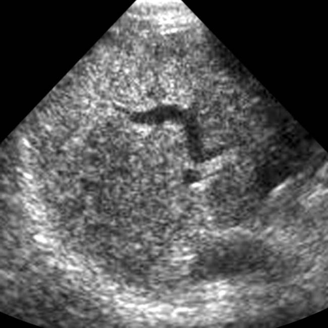
 , while there are innumerable tiny echogenic foci
  throughout the liver that correspond to the smaller biliary hamartomas, which presumably contain relatively more fibrous tissue and less fluid.
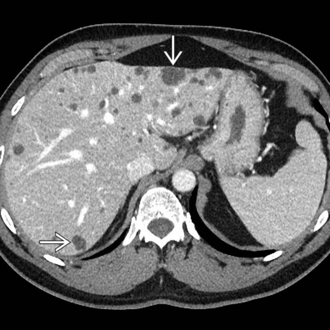
 . These are typical findings for biliary hamartomas and would be a very rare (unreported) feature of hepatocellular carcinoma (HCC).
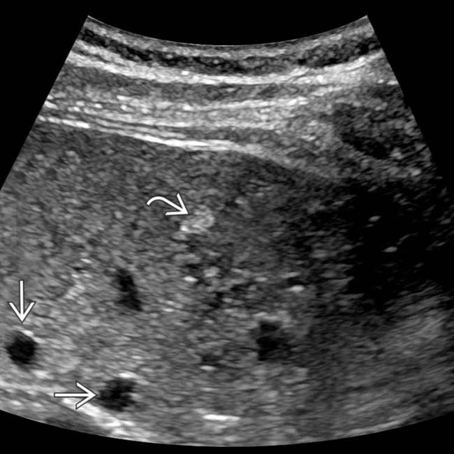
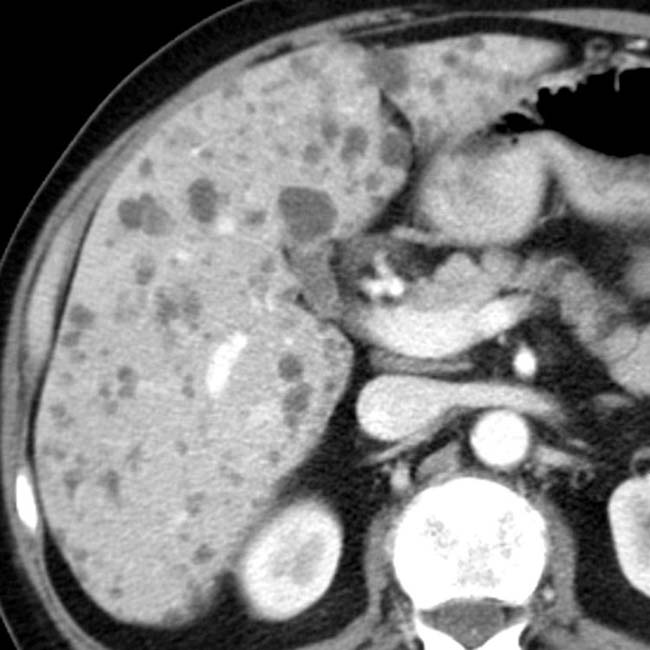
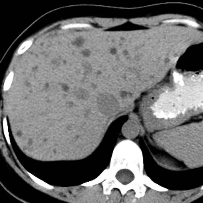
 and many more cystic lesions
  . None of these lesions showed interval change on subsequent imaging studies over several years and likely represent biliary hamartomas.
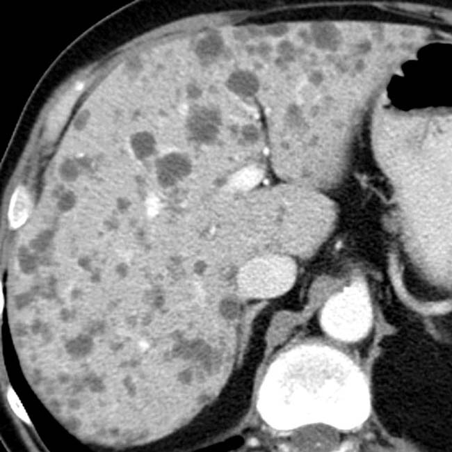
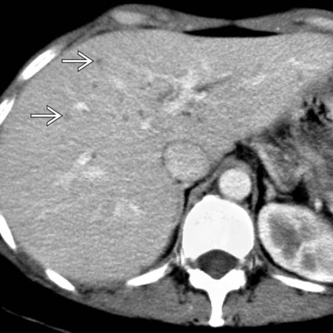
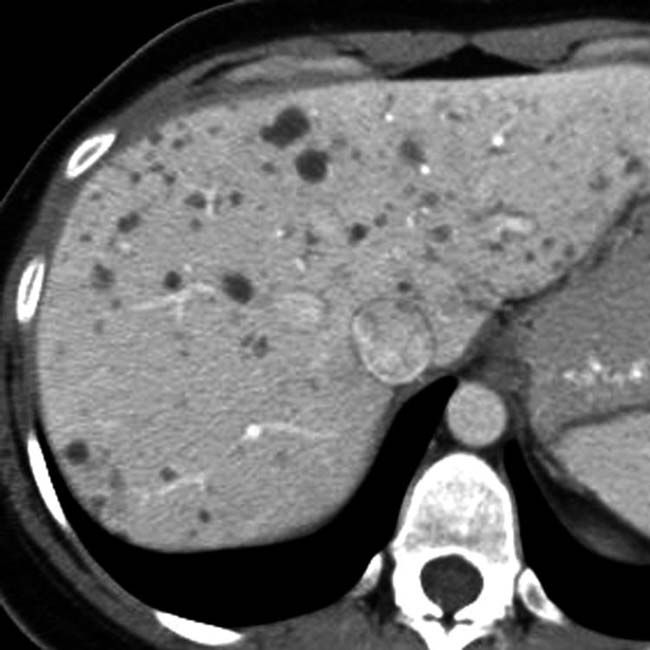
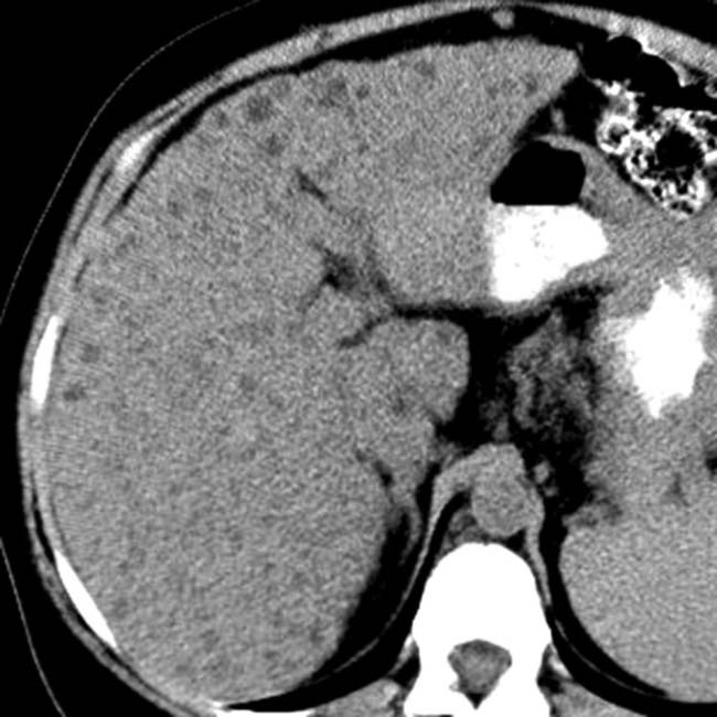
 throughout the liver, too small to permit accurate density measurements. In a healthy, nononcologic patient, they likely represent biliary hamartomas.
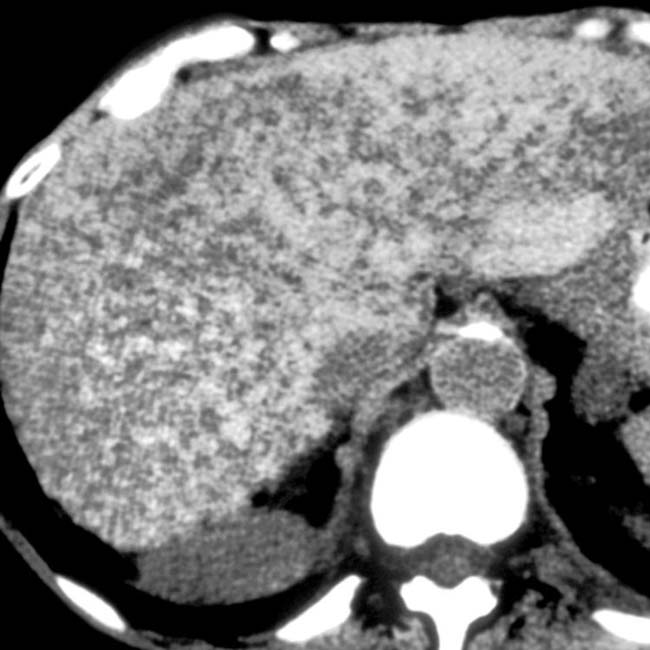
 . This patient also had an enlarged, dysmorphic liver and supernumerary and enlarged hepatic arteries as signs of congenital hepatic fibrosis.
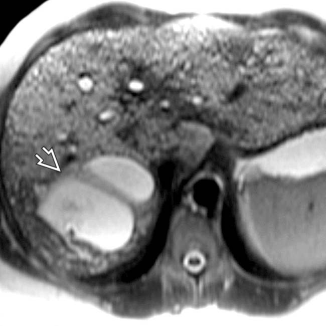
 that communicate with the biliary tree and represent aberrant bile duct cysts of Caroli disease. Congenital hepatic fibrosis, Caroli disease, and biliary hamartomas are all manifestations of fibropolycystic disease.
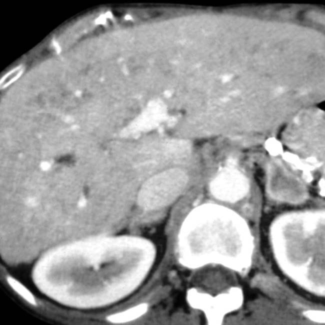
[/level-membership-for-radiology-category][not-level-membership-for-radiology-category] a.k.a. von Meyenburg complex
, representing biliary hamartomas. This patient also had evidence of congenital hepatic fibrosis on imaging and liver biopsy, both part of the congenital hepatic and renal fibropolycystic disease spectrum. that do not communicate with the (normal) biliary tree. This feature helps to distinguish biliary hamartomas from Caroli disease. throughout the liver and 1 of ∼ 10 cyst-like lesions , though even these have small foci of echogenicity within the wall. MR on this patient showed many more cystic-appearing biliary hamartomas.IMAGING
General Features
Angiographic Findings
Buy Membership for Radiology Category to continue reading. Learn more here
[/not-level-membership-for-radiology-category]
