[level-membership-for-radiology-category]
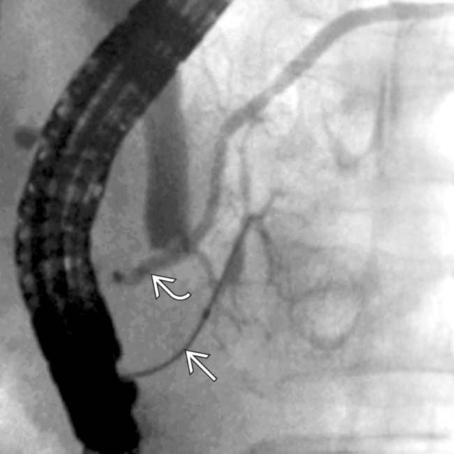
 and accessory
  pancreatic ducts do not communicate. This results embryologically from failure of fusion of the ducts between the dorsal and ventral pancreatic anlagen.
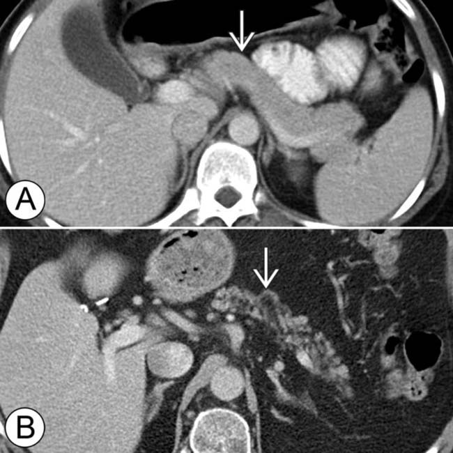
 . The top image (A) is from a 30-year-old woman and the bottom image (B) is from a 78-year-old man. With aging, the pancreas decreases in size with increased fatty lobulation. Small calcifications and mild ductal dilatation may also be seen.
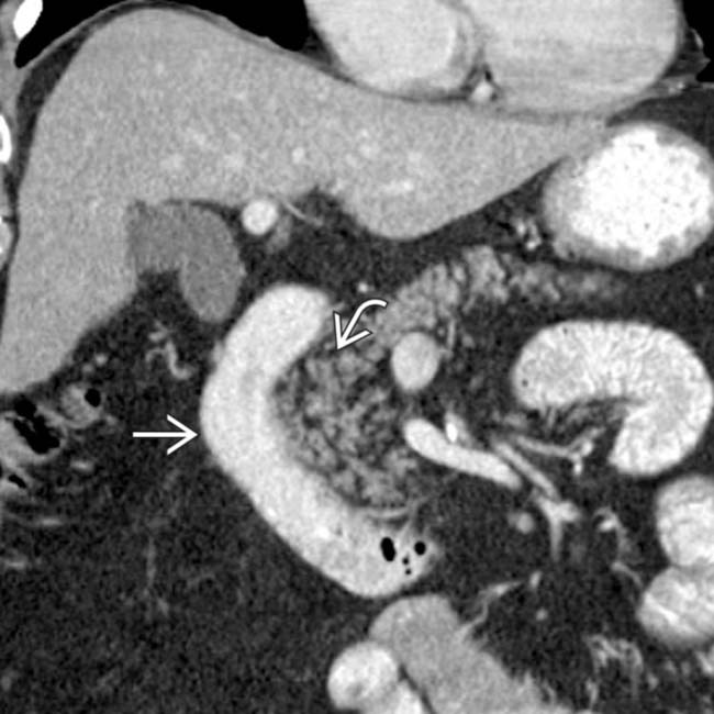
 to the 2nd portion of the duodenum
  .
.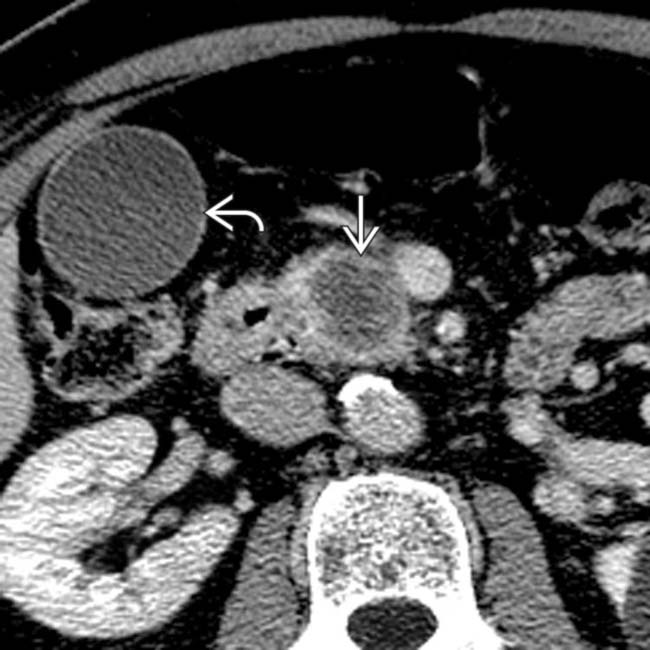
 in the head of the pancreas and a distended gallbladder
  .
.
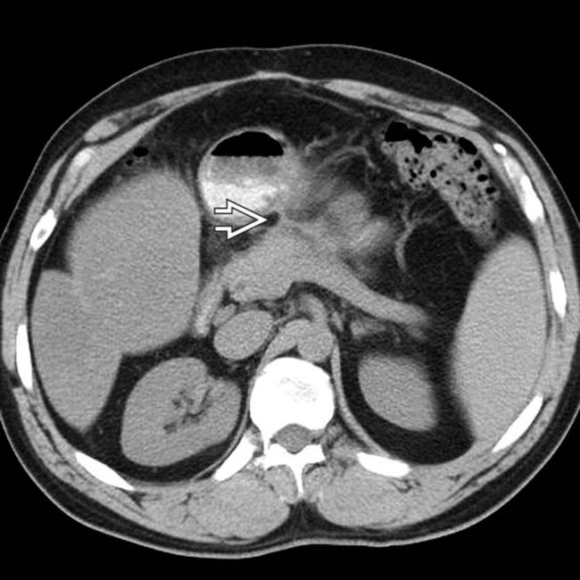
 interrupted as it enters the hypodense mass
  , a typical presentation of pancreatic ductal carcinoma.
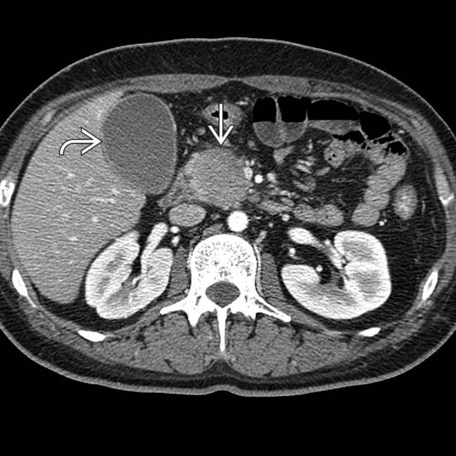
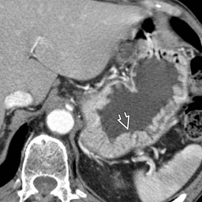
 in the head of the pancreas causing biliary obstruction and dilation of the gallbladder
  .
.
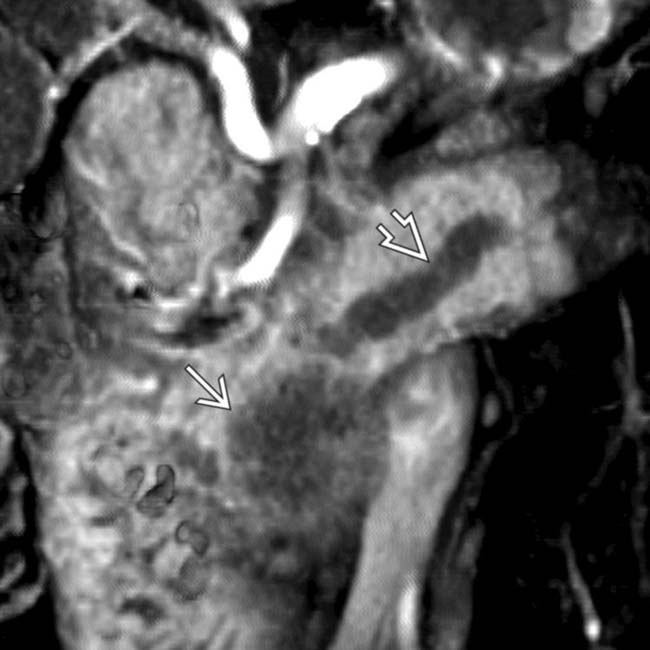
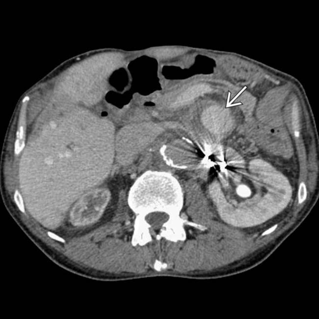
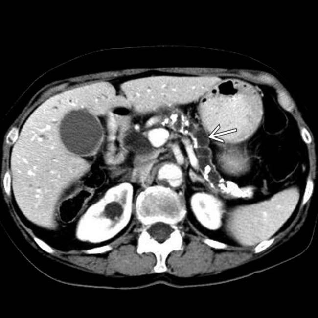
 causing partial obstruction of the bile duct
  , while the pancreatic duct
  is only mildly dilated. Further evaluation, including biopsy, confirmed a diagnosis of autoimmune (IgG4-related) pancreatitis.
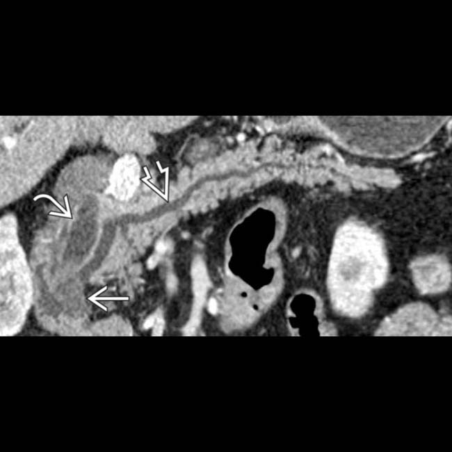
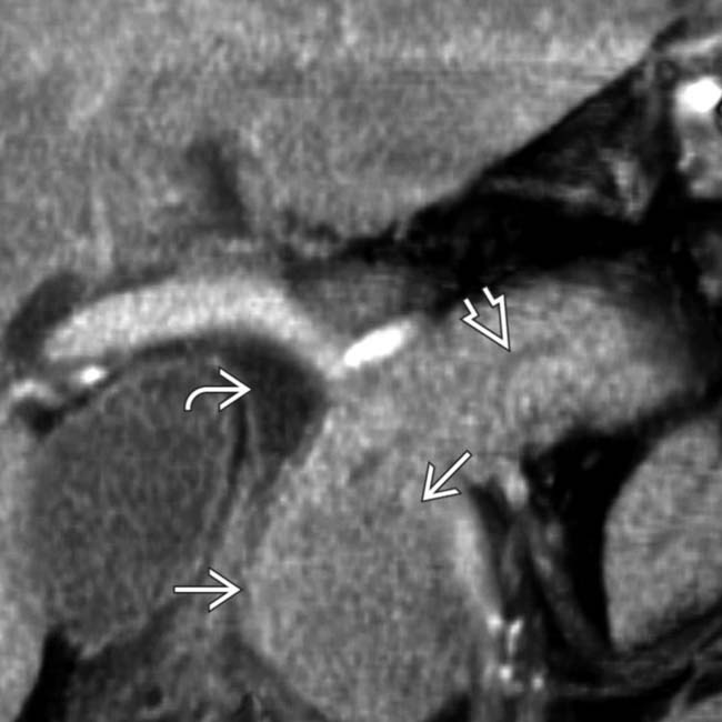
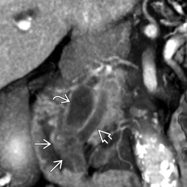
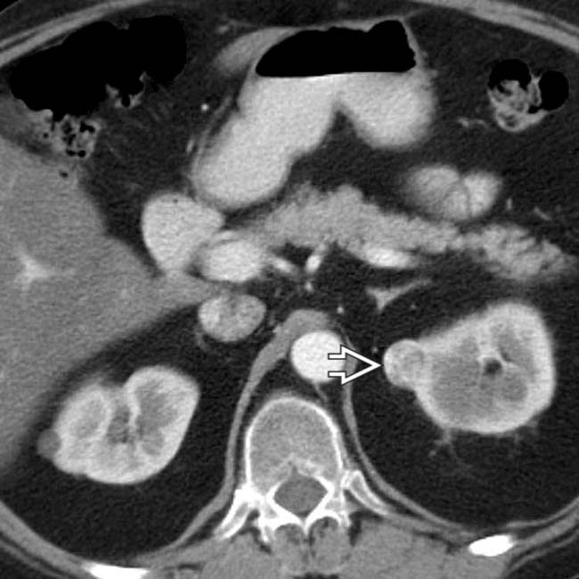
 and pancreatic duct
  due to a small hypodense ampullary carcinoma
  .
.
 , pancreatic duct
  , and ampullary tumor
  .
.
 .
.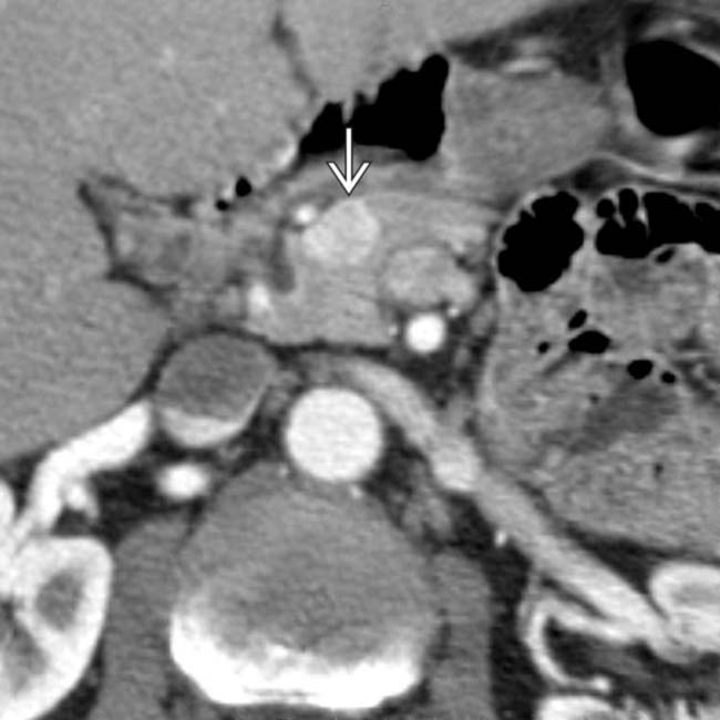
 in the pancreatic head that proved to be a gastrinoma (1 type of pancreatic endocrine tumor) that was responsible for this patient’s Zollinger-Ellison syndrome.
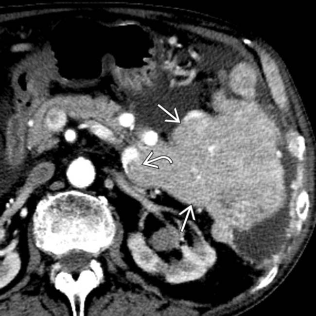
 originating from the pancreas with direct invasion of the splenic vein
  , typical features of a malignant neuroendocrine tumor.
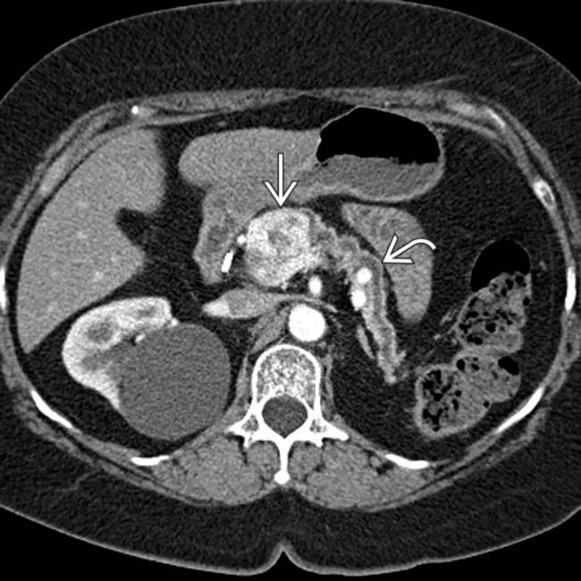
 with parenchymal atrophy and pancreatic ductal dilation upstream
  .
.
 that simulates a hypervascular pancreatic mass.
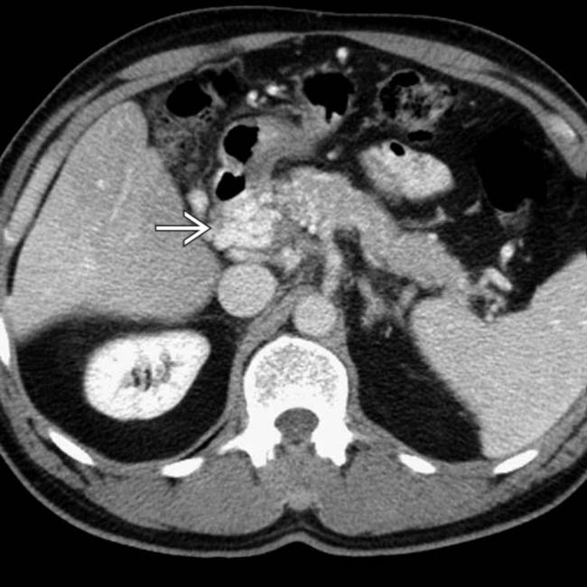
 in and around the pancreatic head that might be mistaken for a hypervascular tumor of the pancreas.
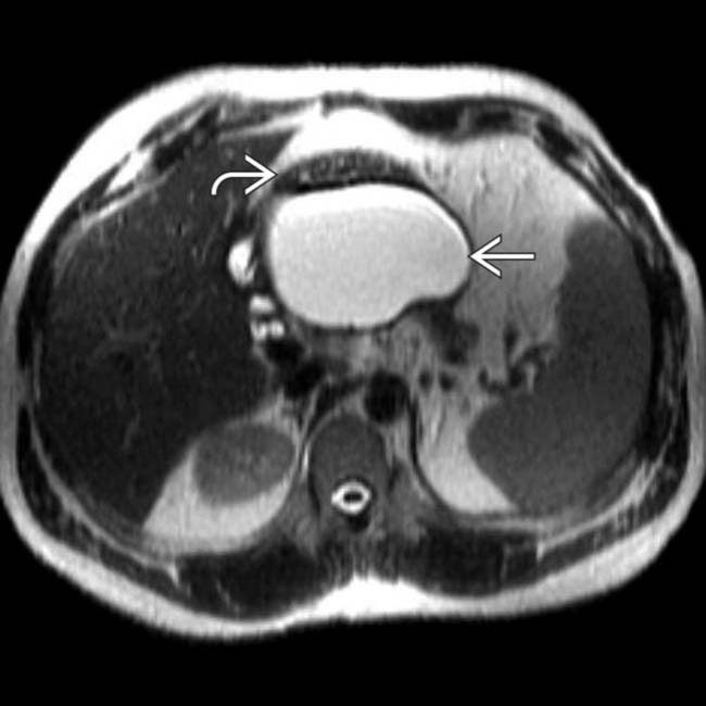
 that displaces the stomach
  .
.
 . The patient’s clinical symptoms improved at the same time. Pseudocysts, unlike cystic pancreatic neoplasms, usually evolve in size quickly, which may aid in distinction.
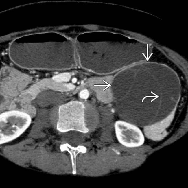
 within the body-tail of the pancreas. Note the septa
  dividing it into several noncommunicating cystic spaces, a classic presentation for mucinous cystic neoplasm.
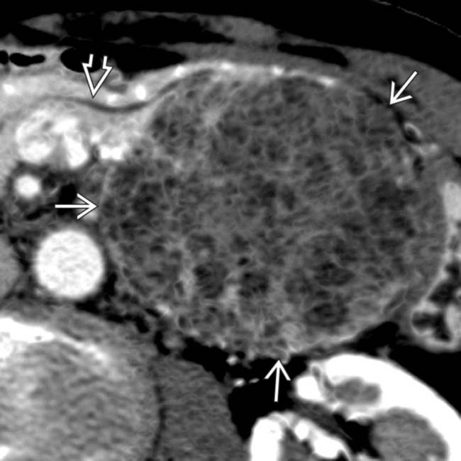
 that displaces, but does not involve, the pancreatic duct
  . The mass has a “honeycomb” or “sponge” appearance, characteristic of serous microcystic adenoma.
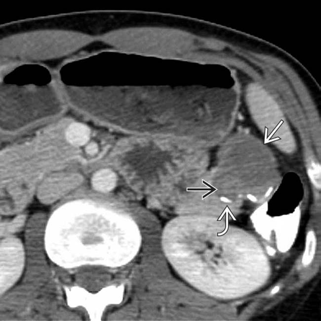
 in the tail of the pancreas, with peripheral rim calcification
  and solid mural nodularity
  , typical features of a solid and papillary epithelial neoplasm (SPEN) of the pancreas.
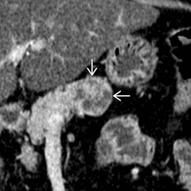
 that proved to be a neuroendocrine tumor.
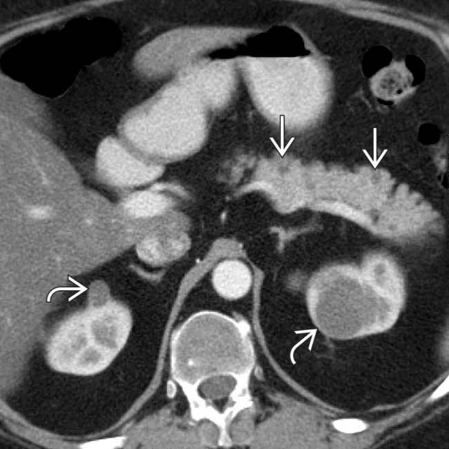
 in the pancreatic body and tail as well as simple cysts in both kidneys
  .
.
 , representing renal cell carcinoma.
 that abuts the pancreas, duodenum, and portal vein
  . The mass is not in the pancreas but is peripancreatic and is a lymphangioma.
 in or near the pancreas that might be mistaken for a cystic pancreatic neoplasm. This represents an aneurysmally dilated and thrombosed portal vein varix in a patient with severe cirrhosis and portal hypertension.
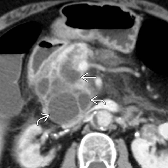
 throughout its length with cystic “masses” in communication with the main and side branches
  .
.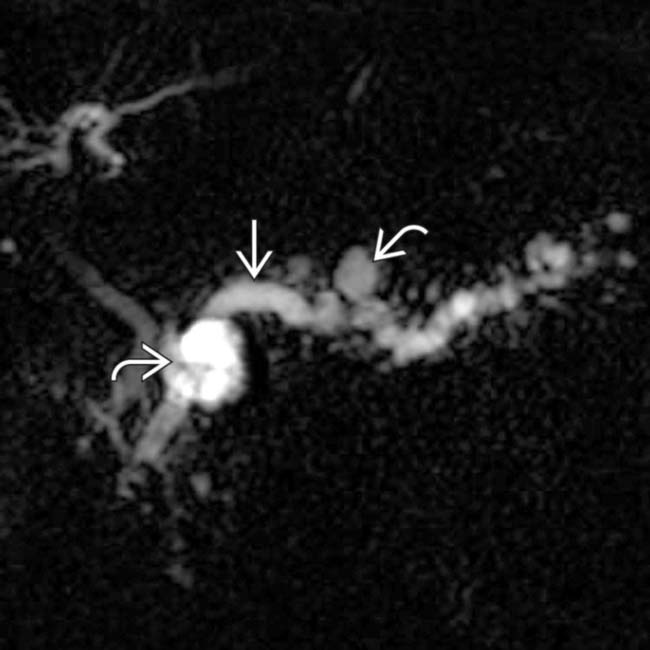
 as well as the side branches
  . This appearance could be due to chronic pancreatitis, but main and side-branch IPMN was confirmed on endoscopic sonography.
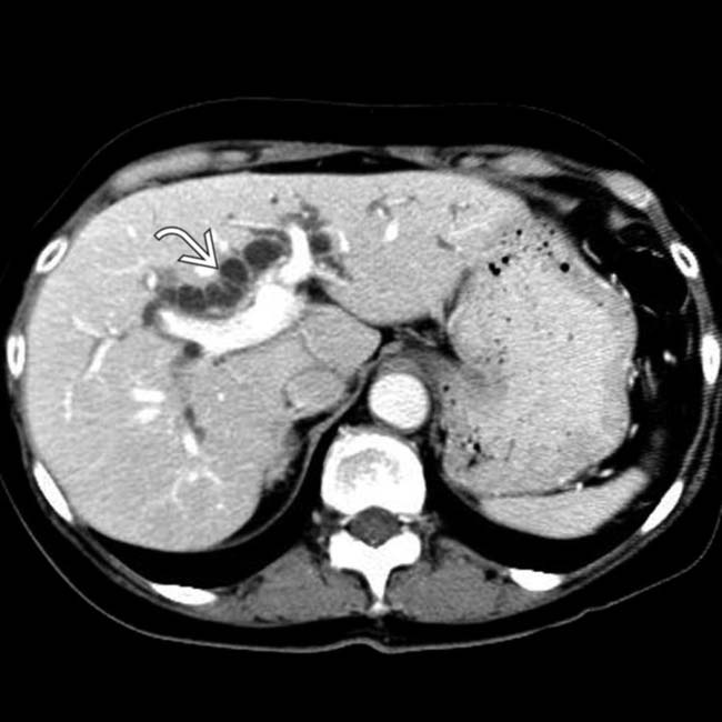
 .
.
 , with extensive calcifications within the ducts and parenchyma. No soft tissue mass was seen and all findings were attributed to chronic pancreatitis.
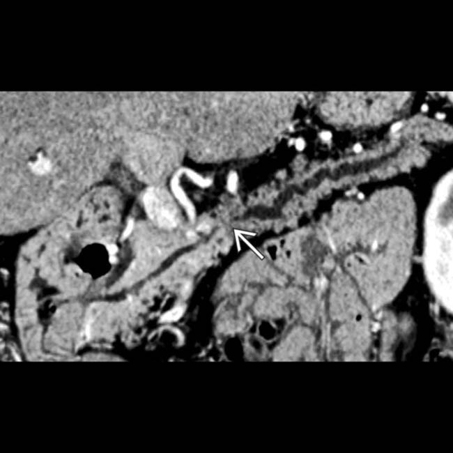
 caused by a small, subtle pancreatic ductal cancer. Multiplanar imaging greatly enhances the ability to evaluate pancreatic masses.
 within the body-tail segment, tapering gradually to a normal caliber duct in the neck. ERCP with sampling of the mucinous pancreatic duct contents confirmed main duct IPMN.
 within the pancreas that are separate from vascular calcifications. There is no pancreatic ductal dilation and these are considered normal senescent changes.
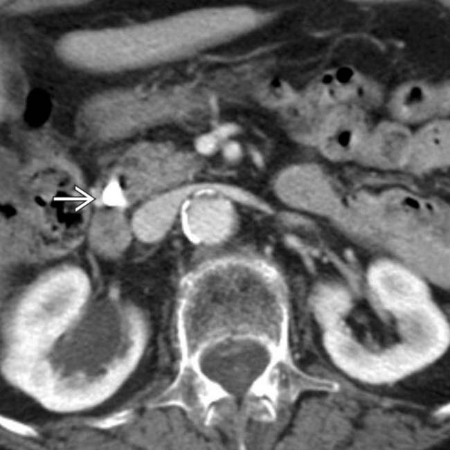
 that is somewhat larger than usually seen in senescent change of the pancreas, but the patient remained symptom-free.
[/level-membership-for-radiology-category][not-level-membership-for-radiology-category]
and accessory pancreatic ducts do not communicate. This results embryologically from failure of fusion of the ducts between the dorsal and ventral pancreatic anlagen.. The top image (A) is from a 30-year-old woman and the bottom image (B) is from a 78-year-old man. With aging, the pancreas decreases in size with increased fatty lobulation. Small calcifications and mild ductal dilatation may also be seen. to the 2nd portion of the duodenum . in the head of the pancreas and a distended gallbladder . interrupted as it enters the hypodense mass , a typical presentation of pancreatic ductal carcinoma. in the head of the pancreas causing biliary obstruction and dilation of the gallbladder . causing partial obstruction of the bile duct , while the pancreatic duct is only mildly dilated. Further evaluation, including biopsy, confirmed a diagnosis of autoimmune (IgG4-related) pancreatitis. and pancreatic duct due to a small hypodense ampullary carcinoma ., pancreatic duct , and ampullary tumor ..Buy Membership for Radiology Category to continue reading. Learn more here
[/not-level-membership-for-radiology-category]
