[level-membership-for-radiology-category] Due to more severe reflux disease


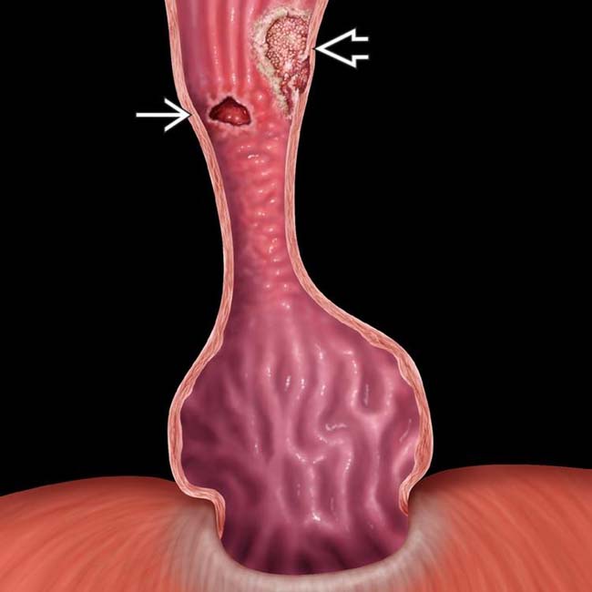
 and an adenocarcinoma
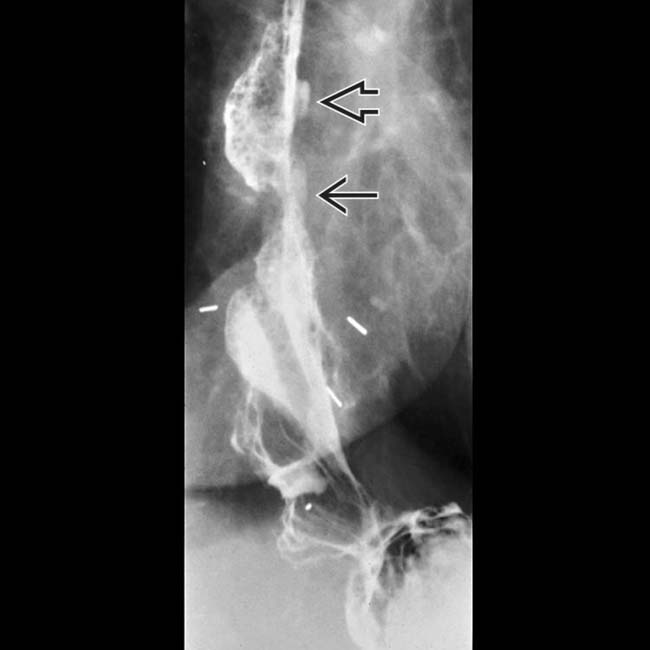
  represented by a raised sessile lesion with an irregular surface.
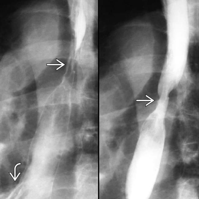
 and ulcer in a patient with a small hernia
  and reflux.
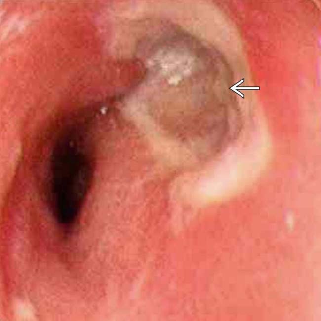
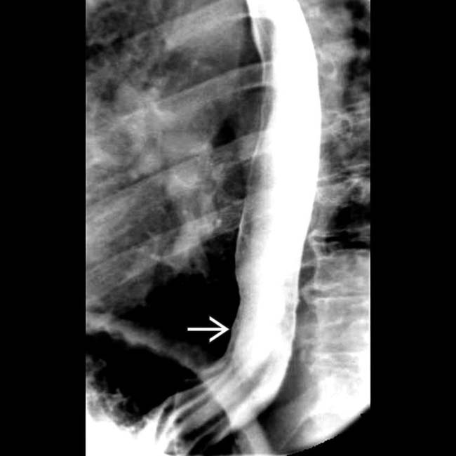
 with the velvet texture of Barrett mucosa and stricture. Normal esophageal mucosa has a shiny, smooth, pink surface.
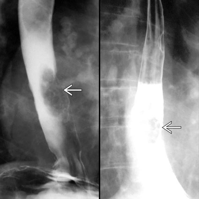
 that represents an adenocarcinoma arising in Barrett mucosa.


 and higher. Note the discrete ulcer
  .
.
 in the distal esophagus in this patient with adenocarcinoma on Barrett mucosa.
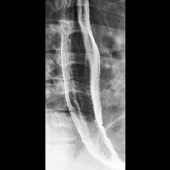
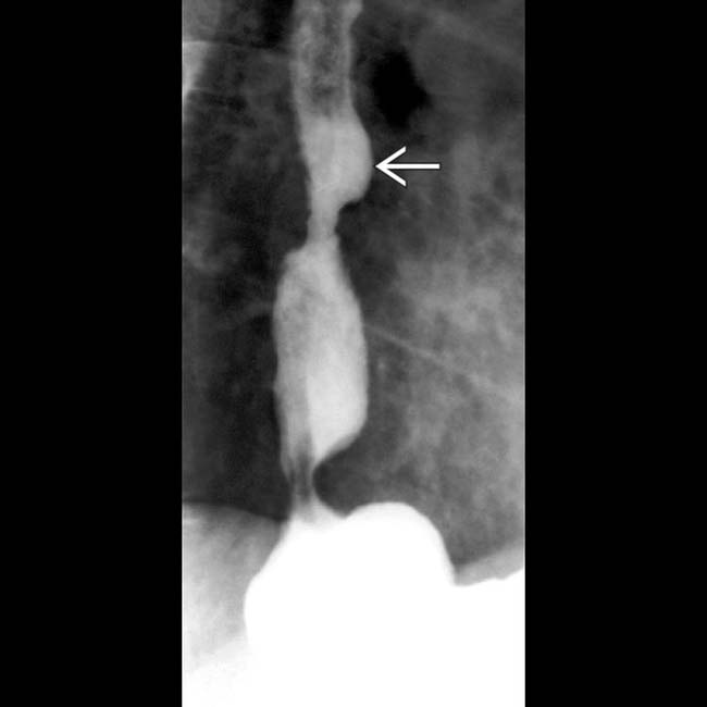
 and hiatal hernia.
[/level-membership-for-radiology-category][not-level-membership-for-radiology-category] Due to more severe reflux disease
and an adenocarcinoma represented by a raised sessile lesion with an irregular surface. and ulcer in a patient with a small hernia and reflux. with the velvet texture of Barrett mucosa and stricture. Normal esophageal mucosa has a shiny, smooth, pink surface. that represents an adenocarcinoma arising in Barrett mucosa.IMAGING
General Features
Radiographic Findings
• Classified into 2 types based on endoscopy and histopathologic findings
 Long segment: Columnar epithelium > 3 cm above gastroesophageal (GE) junction
Long segment: Columnar epithelium > 3 cm above gastroesophageal (GE) junction
Buy Membership for Radiology Category to continue reading. Learn more here
[/not-level-membership-for-radiology-category]
