[level-membership-for-obstetrics-gynecology-category]
Lymph Nodes, Enlarged
Synonyms/Description
Lymphadenopathy
Etiology
Lymph nodes are common sites of metastatic disease in gynecologic tumors and are an important prognostic factor in these malignancies. For example, the 5-year survival for a patient with vulvar cancer and normal nodes is 90%, compared with a patient with nodal disease, whose 5-year survival rate is 50%.
Ultrasound Findings
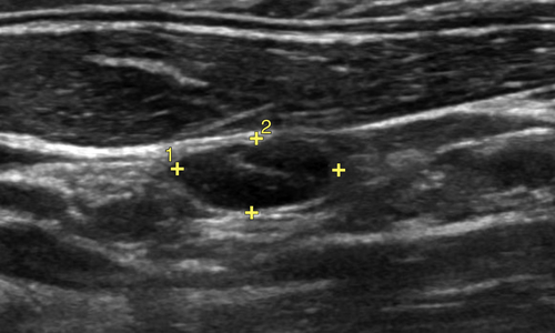
Normal lymph nodes (including pelvic) are typically oblong bean-shaped and small, with the transverse diameter less than or equal to 10 mm. They have a peripheral hypoechogenic band with a hyperechogenic (fatty) hilum. The vascular pattern of normal lymph nodes is characteristic, with the feeding artery and vein coursing in and out from the hilum. Lymph nodes containing tumor tend to be enlarged, with an irregular border and loss of normal sonographic architecture. They are rounded in shape rather than oblong, and their blood-flow pattern can become multifocal and disorganized.
Differential Diagnosis
Sonographically, an enlarged lymph node appears as a mass, and may be difficult to distinguish from any other solid mass in the pelvis, unless the location and pattern of blood flow suggest a lymph node. The color flow pattern of a lymph node will virtually always have a single vascular source.
Clinical Aspects and Recommendations
Ultrasound may be the initial method of detection of abnormal pelvic lymph nodes. This finding carries important prognostic and therapeutic implications and may help to stage disease in a cancer patient. PET/CT and other imaging techniques are likely to be used to further determine extent of disease and treatment.
Figures
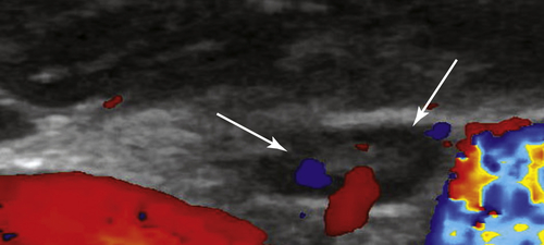
Figure L1-1 Normal lymph node. The normal lymph node is identified by calipers. Color flow to the node (arrows) shows a single source of vessels along the long axis of the small node.
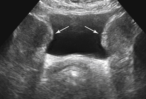
Figure L1-2 Patient with chronic lymphocytic leukemia. Transverse view of the pelvis in a patient with a full bladder showing bilateral solid masses along the pelvic side walls indenting the sides of the bladder.
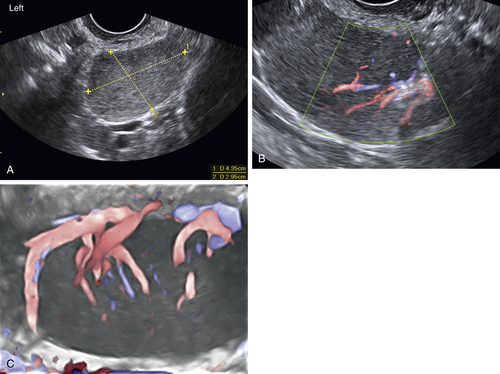
Figure L1-3 A, Magnified view of a left-sided mass seen transvaginally, showing a homogeneously solid mass (calipers). This enlarged and abnormal node is rounded and wide, with loss of normal architecture. B, The same mass is interrogated with color flow Doppler, showing that the blood flow into the mass originates from one side, characteristic of a lymph node. C, The contralateral lymph node (seen here using 3-D color Doppler) is also enlarged with abundant color flow.
Suggested Reading
Fischerova D. Ultrasound scanning of the pelvis and abdomen for staging of gynecological tumors: a review. Ultrasound Obstet Gynecol. 2011;38:246–266.
Lai G., Rockall A.G. Lymph node imaging in gynecologic malignancy. Semin Ultrasound CT MRI. 2010;31:363–376.
Gore R.M., Newmark G.M., Thakrar K.H., Mehta U.K., Berlin J.W. Pelvic incidentalomas. Cancer Imaging. 2010;10:S15–S26.
[/level-membership-for-obstetrics-gynecology-category][not-level-membership-for-obstetrics-gynecology-category]
Lymph Nodes, Enlarged
Synonyms/Description
Lymphadenopathy
Etiology
Lymph nodes are common sites of metastatic disease in gynecologic tumors and are an important prognostic factor in these malignancies. For example, the 5-year survival for a patient with vulvar cancer and normal nodes is 90%, compared with a patient with nodal disease, whose 5-year survival rate is 50%.
Ultrasound Findings
Normal lymph nodes (including pelvic) are typically oblong bean-shaped and small, with the transverse diameter less than or equal to 10 mm. They have a peripheral hypoechogenic band with a hyperechogenic (fatty) hilum. The vascular pattern of normal lymph nodes is characteristic, with the feeding artery and vein coursing in and out from the hilum. Lymph nodes containing tumor tend to be enlarged, with an irregular border and loss of normal sonographic architecture. They are rounded in shape rather than oblong, and their blood-flow pattern can become multifocal and disorganized.
Differential Diagnosis
Buy Membership for Obstetrics & Gynecology Category to continue reading.
Learn more here
[/not-level-membership-for-obstetrics-gynecology-category]
Gynecologic Ultrasound A Problem-Based Approach Expert Consult