[level-membership-for-internal-medicine-category]
CASE 50 Renal Artery Stenosis Resulting From Fibromuscular Dysplasia
Cardiac catheterization
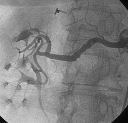
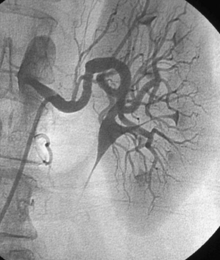
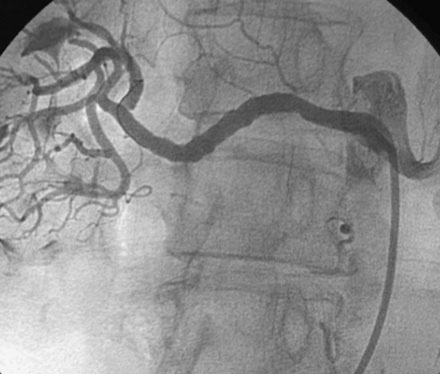
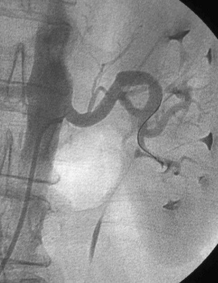
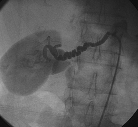
A diagnostic cardiac catheterization was performed first and revealed mild coronary atherosclerosis with no significant obstruction and preserved left ventricular function. Selective right renal angiography demonstrated the characteristic findings of fibromuscular dysplasia with a “beaded” appearance of the renal artery and the presence of linear “webs” (Figure 50-1 and Video 50-1). This abnormality was also present to a lesser degree in the left renal artery (Figure 50-2 and Video 50-2). After administering 70 U/kg of unfractionated heparin, balloon dilatation was performed at these sites using a 5.5 mm diameter by 20 mm long balloon, which improved the angiographic appearance (Figures 50-3, 50-4 and Videos 50-3, 50-4).
Discussion
Renal artery stenosis is most commonly caused by atherosclerosis, typically affecting the ostium and proximal segment of the renal artery. In roughly 10% of cases, fibromuscular dysplasia is the cause.1 This condition is more common in young females (less than 50 years old) and involves the distal portion of the renal artery or its branches. Although usually attributed to young women, it has also been recognized as a cause of secondary hypertension in elderly individuals, as shown in this case.2
The diagnosis of fibromuscular dysplasia is made based on the classic angiographic appearance, often referred to as a “string of beads” (Figure 50-5). This finding is due to luminal occupation with webs and ridges of dysplastic media. It is sometimes difficult to decide if these webs and ridges obstruct the lumen to a significant degree. When the angiogram is ambiguous, additional information regarding the hemodynamic significance of an abnormal segment can be determined by measurement of a pressure gradient.3
In general, fibromuscular dysplasia is treated with balloon angioplasty alone. Disruption of the abnormal webs and ridges with a balloon alone is often adequate and stenting is reserved for treatment of flow-limiting dissections caused by the balloon. The clinical response to balloon angioplasty is excellent, with more than 90% cured or improved after the procedure and restenosis occurring in about 20%.4
1 Safian R.D., Textor S.C. Renal artery stenosis. N Engl J Med. 2001;344:431-442.
2 Pascual A, Bush HS, Copley JB: Renal fibromuscular dysplasia in elderly persons, Am J Kidney Dis 45:E63–E66
3 Birrer M., Do D.D., Mahler F., et al. Treatment of renal artery fibromuscular dysplasia with balloon angioplasty: A prospective follow-up study. Eur J Vasc Endovasc Surg. 2002;23:146-152.
4 Mahmud E., Brocato M., Palakodeti V., et al. Fibromuscular dysplasia of renal arteries: Percutaneous revascularization based on hemodynamic assessment with a pressure measurement guidewire. Catheter Cardiovasc Interv. 2006;67:434-437.
[/level-membership-for-internal-medicine-category][not-level-membership-for-internal-medicine-category]
CASE 50 Renal Artery Stenosis Resulting From Fibromuscular Dysplasia
Cardiac catheterization
A diagnostic cardiac catheterization was performed first and revealed mild coronary atherosclerosis with no significant obstruction and preserved left ventricular function. Selective right renal angiography demonstrated the characteristic findings of fibromuscular dysplasia with a “beaded” appearance of the renal artery and the presence of linear “webs” (Figure 50-1 and Video 50-1). This abnormality was also present to a lesser degree in the left renal artery (Figure 50-2 and Video 50-2). After administering 70 U/kg of unfractionated heparin, balloon dilatation was performed at these sites using a 5.5 mm diameter by 20 mm long balloon, which improved the angiographic appearance (Figures 50-3, 50-4 and Videos 50-3, 50-4).
