CASE 200
History: A 50-year-old woman presents with upper abdominal pain.
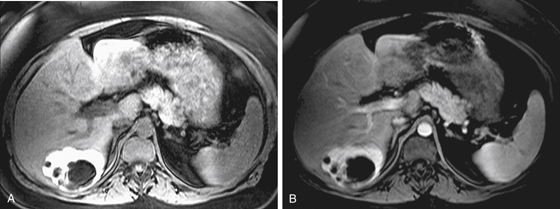
1. What should be included in the differential diagnosis of the imaging finding shown in Figure A? (Choose all that apply.)
2. Which of the following statements regarding biliary cystadenomas is true?
A. Biliary cystadenomas are more common in women.
B. The most common presentation is jaundice.
C. Most progress to cystadenocarcinoma.
D. The treatment of choice is enucleation of the cyst.
3. Which of the following imaging features is most useful to differentiate a biliary cystic neoplasm from a simple hepatic cyst?
4. What might ultrasound show in a biliary cystadenoma that is not evident in these MRI images and might not be seen with CT?
ANSWERS
CASE 200
Cystadenoma of the Biliary Ducts
1. A, C, and E
2. A
3. C
4. A
References
Kim JY, Kim SH, Eun HW, et al: Differentiation between biliary cystic neoplasms and simple cysts of the liver: accuracy of CT. AJR Am J Roentgenol. 2010;195(5):1142-1148.
Cross-Reference
Gastrointestinal Imaging: THE REQUISITES, 3rd ed, p 203.
Comment
Biliary cystadenomas are rare cystic lesions arising from the bile ducts (see figures). They are most commonly seen in middle-aged women and have some malignant potential. Patients often present with upper abdominal discomfort; rarely, jaundice or mass effect is the presentation. The finding may be incidental most of the time. The lesions are multiloculated and septated, and this is usually more apparent with ultrasound than with CT or MRI.
Because this lesion has malignant potential, and diagnostic imaging is unable to differentiate between a cystadenoma and a cystadenocarcinoma, needle aspiration biopsy or surgical removal is usually required for definitive histologic diagnosis. Both are slow-growing, and both can have similar imaging appearances. The lesions can be of varying sizes, with some 3 to 5 cm and others 30 cm. Alternative diagnostic considerations include hepatic abscess and congenital biliary cysts. The latter are usually multiple, and biliary cystadenomas are usually solitary. In the array of cystic lesions that may be seen in the liver, cystadenomas must be considered relatively rare, accounting for 1% to 5% of all hepatic cystic processes. In some reported cases, symptomatic patients with a cystadenoma were misdiagnosed (i.e., a hydatid cyst). Generally, when a multiloculated septated cyst (usually in the right lobe of the liver) is discovered in a middle-aged woman, biliary cystadenoma or cystadenocarcinoma should be considered in the differential diagnosis.


