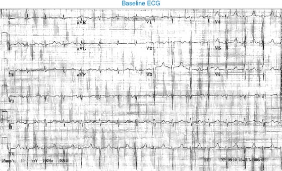
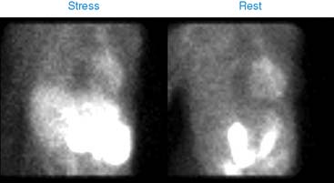
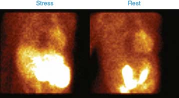
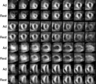
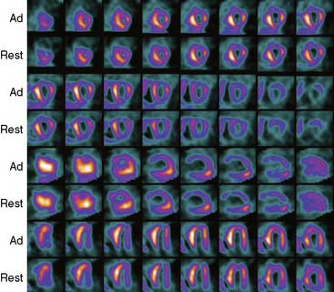
Case 20
1. Williams K.A., Hill K.A., Sheridan C.M. Noncardiac findings on dual-isotope myocardial perfusion SPECT. J Nucl Cardiol. 2003;10:395-402.
2. Raza M., Meesala M., Panjrath G., et al. Abnormal photopenic area on nuclear perfusion imaging. J Nucl Cardiol. 2005;12:607-609.
3. Raza M., Panjrath G., Jain D. Unusual retrocardiac radiotracer uptake on sestamibi perfusion images. J Nucl Cardiol. 2004;11:e1-e2.
4. Meesala M., Raza M., Yaganti V., et al. Bilateral photopenic areas in the lungs on SPECT imaging. J Nucl Cardiol. 2006;13:728-730.
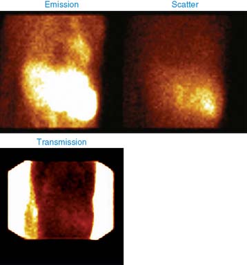
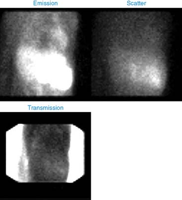
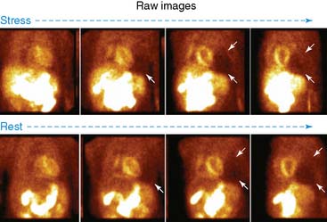
5. Hendel R.C., Gibbons R.J., Bateman T.M. Use of rotating (cine) planar images in the interpretation of a tomographic myocardial perfusion study. J Nucl Cardiol. 1999;6:234-240.
6. Panjrath G.S., Narra K., Jain D. Myocardial perfusion imaging in a patient with chest pain. J Nucl Cardiol. 2004;11:515-517.


