CASE 2
1. What should be included in the differential diagnosis for mitral regurgitation? (Choose all that apply.)
D. Endocarditis
2. What is the most likely diagnosis?
D. Endocarditis
3. Which imaging plane is ideal for diagnosing this condition?
A. Four-chamber
B. Short-axis
C. Left ventricular outflow tract (3-chamber)
D. Right ventricular outflow tract
4. Which of the following is a potential complication of this condition?
C. Arrhythmia
ANSWERS
References
Feuchtner GM, Alkadhi H, Karlo C, et al. Cardiac CT angiography for the diagnosis of mitral valve prolapse: comparison with echocardiography. Radiology. 2010;254(2):374–383.
Han Y, Peters DC, Salton CJ, et al. Cardiovascular magnetic resonance characterization of mitral valve prolapse. JACC Cardiovasc Imaging. 2008;1(3):294–303.
Cross-Reference
Cardiac Imaging: The REQUISITES, ed 3, pp 192–193.
Comment
Epidemiology
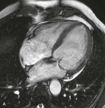
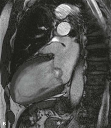
Mitral valve prolapse affects 2% to 3% of the population and is the most common cause of severe nonischemic mitral regurgitation requiring surgery (Fig. A). It is defined by greater than 2 mm of atrial displacement of the valve leaflets (measured on left ventricular outflow tract or vertical long-axis [2-chamber] images) with respect to the mitral anulus (Fig. B). There are two types of mitral valve prolapse: billowing (as shown in this case) and flail leaflet (caused by chordal rupture secondary to endocarditis or rheumatic heart disease). Mitral valve prolapse is most commonly due to myxomatous degeneration of mitral leaflets but can also be seen in patients with atrial secundum septal defects, connective tissue disorders, and recent diuretic use.
Imaging
Mitral valve prolapse may be incorrectly diagnosed if only four-chamber images are viewed secondary to the natural saddle shape of the mitral anulus. Myxomatous leaflet (defined as a leaflet thickness measuring >5 mm) predisposes patients to fatal arrhythmia, severe mitral regurgitation, and endocarditis. It is important to identify mitral valve prolapse on cardiac CT or MRI examinations performed for other reasons because these patients may benefit from endocarditis prophylaxis or anticoagulation therapy.