CASE 199
History: A 44-year-old woman presents with abdominal pain.
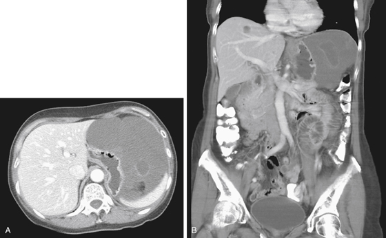
1. What should be included in the differential diagnosis of the imaging finding shown in Figure A? (Choose all that apply.)
A. Subphrenic fluid collection
2. Where are gastrointestinal stromal tumors (GISTs) most commonly found?
3. What is the most common imaging morphology of a GIST?
C. Intramural, submucosal mass
D. Exophytic, extragastric extension
4. What is the most common cause of a bulky gastric tumor?
ANSWERS
CASE 199
Cystic Gastric GIST
1. A, B, C, and D
2. C
3. D
4. A
References
Darnell A, Dalmau E, Pericay C, et al: Gastrointestinal stromal tumors. Abdom Imaging. 2006;31(4):387-399.
Cross-Reference
Gastrointestinal Imaging: THE REQUISITES, 3rd ed, p 77.
Comment
Many GISTs encountered in the bowel and particularly the stomach behave in a benign fashion. They may be ulcerated and bleed if in the wall of the bowel. Most are solid tumors, although some show areas of tumor necrosis and liquefaction and resultant mixed density. The GIST in this case shown on CT is problematic because it does not seem to be “arising” from any particular organ and is almost entirely cystic, with the exception of some foci of density seen on the axial view (see figures). Initially, GISTs were considered mesenchymal or smooth muscle tumors, leiomyomas, or leiomyosarcoma. However, further histologic investigation suggested a surprising lack of smooth muscle elements in the lesion. As a result, the term gastrointestinal stromal cell tumor was introduced to denote more correctly the spindle cell and epithelioid elements that compose these tumors.
The tumor can undergo considerable necrosis and manifest as near-cystic lesions in some cases. However, there is also a distinctive morphologic variant of characterized by myxoid content that is well encapsulated, cystic in appearance, mostly seen arising exophytically from the stomach, and mostly seen in women, and such a lesion is shown in this case.


