CASE 189
History: A 19-year-old man presents with right costal margin pain and swelling.
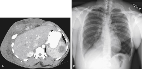
1. What should be included in the differential diagnosis of the imaging finding shown in Figure A? (Choose all that apply.)
2. Which of the following statements regarding cystic splenic lesions is true?
A. Most cystic splenic lesions are neoplastic.
B. Splenic lymphangioma is usually diagnosed in childhood.
C. Patients with echinococccal cysts develop fever secondary to antigen release.
3. What is the most common clinical manifestation of a lymphangioma?
4. Which of the following conditions is associated with lymphangiomas?
ANSWERS
CASE 189
Lymphangioma of the Spleen
1. B, C, D, and E
2. B
3. B
4. D
References
Kamaya A, Weinstein S, Desser TS. Multiple lesions of the spleen: differential diagnosis of cystic and solid lesions. Semin Ultrasound CT MRI. 2006;27(5):389-403.
Warshauer DM, Hall HL: Solitary splenic lesions. Semin Ultrasound CT MRI. 2006;27(5):370-388.
Cross-Reference
Gastrointestinal Imaging: THE REQUISITES, 3rd ed, p 214.
Comment
Lymphangioma is an unusual condition that rarely involves the spleen. The splenic lesions can be solitary or multiple as in this case (see figures). Lymphangioma is a congenital malformation of the lymphatic vessels and system. It can be focal or widespread lymphangiomatosis, as in this case. The lesion is entirely benign with no malignant potential known. Perhaps the most common manifestation in infants is a cystic hygroma, which is thought to represent a form of focal lymphangioma in the neck. Histologically, lymphangiomas are thought to be hamartomas rather than neoplasms.
Splenic involvement can be solitary organ involvement or part of lymphangiomatosis. Bone also can be involved. Deformities of the ribs in this patient are noted (see figures). Lytic expansile lesions of bones in this condition have been called Gorham’s disease. However, these bones appear deformed rather than replaced or destroyed. In some cases of splenic lymphangioma, splenectomy has been necessary when the lesion is symptomatic or diagnosis is questionable. The skin thickening seen over this patient’s body is a manifestation of lymphangiomatosis, as is the mediastinal and neck involvement. This condition has been classified into three groups: (1) lymphangioma circumscriptum, (2) cavernous lymphangioma (this patient), and (3) cystic lymphangioma (cystic hygroma, which can also be seen in some cases of cavernous lymphangioma).


