CASE 187
History: A 57-year-old man presents with vague left upper quadrant pain.
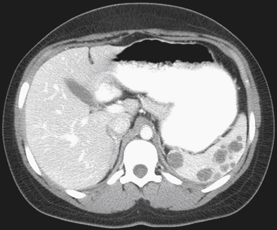
1. What should be included in the differential diagnosis of the imaging finding shown in the figure? (Choose all that apply.)
2. What is the most common cause of perfusion defects in the spleen?
3. What is the most common tumor seen in the spleen?
4. Which of the following statements regarding splenic metastases is true?
A. Splenic involvement is rare in patients with widespread metastases.
B. Calcified splenic metastases arise most commonly from an ovarian primary.
C. The most common primary site of hematogenous metastases is GI tumors.
ANSWERS
CASE 187
Splenic Metastatic Disease
1. A, B, and D
2. A
3. C
4. A
References
Kamaya A, Weinstein S, Desser TS. Multiple lesions of the spleen: differential diagnosis of cystic and solid lesions. Semin Ultrasound CT MRI. 2006;27(5):389-403.
Cross-Reference
Gastrointestinal Imaging: THE REQUISITES, 3rd ed, p 212.
Comment
Marginal perfusion defects in the spleen, often wedge-shaped, are a common finding in the spleen at CT imaging. These defects are almost always old splenic infarcts. Despite the vascularity and the hematogenous “filter” function of the spleen, metastatic disease is rare. Several theories as to why this seemingly unusual situation should be the case have been put forward—ranging from the angle of the splenic artery from its takeoff from the celiac axis to the high concentrations of lymphoid tissue in the spleen. As in this case, the most common primary cancer is lung cancer followed by GI tumors (see figure). Splenic metastatic disease apparently without liver involvement is especially rare, representing less than 5% of cases of reported splenic metastatic disease.
Other differential considerations include congenital and acquired (usually secondary to trauma) splenic cysts. These cysts are almost entirely solitary. Hydatid cyst may also be seen in the spleen as a solitary lesion. Abscesses, which are encountered more frequently in immunocompromised patients, may be single but are usually multiple and often tiny. Hemangiomas, the most common benign tumors of the spleen, can be solitary or multiple. Involvement of the spleen with lymphoma or leukemia usually takes the form of splenomegaly. However, if the spleen is normal in size with multiple defects, these possibilities must also be considered. Pancreatic pseudocysts have been reported as intrasplenic lesions.


