CASE 185
History: A 75-year-old man presents with bright rectal bleeding. Colonoscopy had failed because of an obstructive bowel deformity at the splenic flexure.
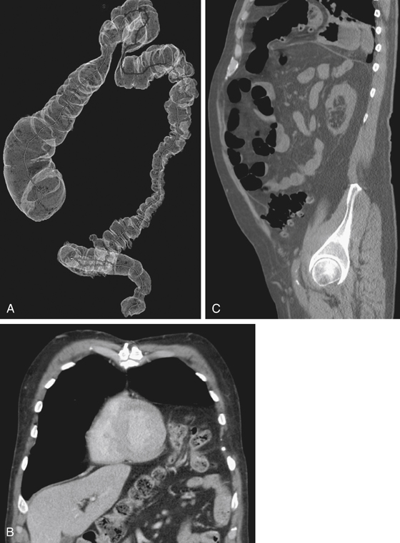
1. What should be included in the differential diagnosis of the imaging finding shown in Figure A? (Choose all that apply.)
A. Traumatic diaphragmatic hernia
2. What is the most common congenital hernia in the diaphragm?
3. Which of the following statements regarding traumatic diaphragmatic hernias is true?
A. Penetrating diaphragmatic injury more commonly occurs on the left side.
B. Iatrogenic diaphragmatic injury is most commonly associated with cardiac surgery.
C. Diaphragmatic injury and hernia caused by blunt force trauma is usually evident at presentation.
4. What is the most common content of Bochdalek’s hernia?
ANSWERS
CASE 185
Diaphragmatic Hernia
1. A, B, and C
2. B
3. A
4. A
References
Eren S, Kantarci M, Okur A. Imaging of diaphragmatic rupture after trauma. Clin Radiol. 2006;61(6):467-477.
Cross-Reference
Gastrointestinal Imaging: THE REQUISITES, 3rd ed, p 326.
Comment
Congenital diaphragmatic hernias are a major concern in neonates and can be associated with other congenital abnormalities, such as pulmonary hypoplasia and gastrointestinal and cardiac abnormalities. The subsequent migration of the abdominal content into the chest can also compromise breathing. Some of these defects may go undetected into adult life and may be discovered as an incidental finding. The same may be true about acquired diaphragmatic hernias following blunt trauma to the abdomen.
With CT, diaphragmatic trauma may be missed at initial imaging, even with multiplanar imaging, if there has been no migration of abdominal content through the rent in the diaphragm. Patients invariably have other life-threatening injuries, which must be addressed first. The left diaphragm is the side most commonly affected with trauma. The liver is thought to confer some protection to the right. The diagnosis is sometimes made at surgery. In many instances in adult life, abdominal content migrates through a congenital or acquired defect in the diaphragm and causes no symptoms. In other patients, there may be vague upper abdominal discomfort. However, some patients eventually develop an incarceration of bowel in the lower chest with obstructive symptoms and potential vascular compromise.
Barium studies have always been a definitive way to evaluate the type and amount of bowel entrapped with the hernia. However, more recently, CT is being used with greater frequency with satisfactory results because of its speed and its ability to evaluate for the presence of other organs besides bowel (spleen and liver) (see figures).


