CASE 184
History: An 18-year-old man presents with a right lower quadrant abdominal mass.
1. What should be included in the differential diagnosis of the imaging finding shown in Figure A? (Choose all that apply.)
2. What is the most common gastrointestinal tract site of involvement by Burkitt’s lymphoma?
3. What is the most common site of presentation of Burkitt’s lymphoma?
4. What virus is specifically associated with Burkitt’s lymphoma?
ANSWERS
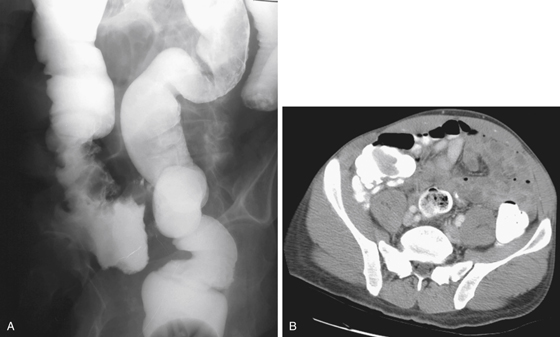
CASE 184
Burkitt’s Lymphoma of the Cecum
1. A, B, C, and E
2. A
3. C
4. C
References
Biko DM, Anupindi SA, Hernandez A, et al: Childhood Burkitt lymphoma: abdominal and pelvic imaging findings. AJR Am J Roentgenol. 2009;192(5):1304-1315.
Cross-Reference
Gastrointestinal Imaging: THE REQUISITES, 3rd ed, p 282.
Comment
This young patient has a destructive colonic mass in the cecum (see figures). The obvious possibility would be colonic adenocarcinoma, which is the most common malignant lesion of the colon. Lymphoma is uncommon. However, among the various types of lymphomas seen in the colon is Burkitt’s lymphoma, named after the pathologist who first described the relationship of this highly aggressive tumor affecting mostly children in equatorial Africa in areas of endemic malaria. The lesion usually involved the mandible in these children.
Burkitt’s lymphoma is one of the most aggressive tumors known, with a doubling rate much greater than the usual adenocarcinoma of the colon. The type found in North America usually manifests as an intra-abdominal process in the distal ileum or proximal colon and can be seen as a mass in the right lower quadrant. It is extremely typical of Burkitt’s lymphoma to involve this area, and the diagnosis should be strongly considered in any child who has a painless palpable mass in the region with heme-positive stool. However, in North America, the disease is seen more commonly in young adults.
In the type of lymphoma that Burkitt described in Africa, the jaw and retroperitoneal lymph nodes were frequently involved in children, but this is not typical of the type of Burkitt’s lymphoma common in North America. The tumor is usually intra-abdominal and often associated with EBV, similar to some other lymphomas and cancers. A distinctive feature of Burkitt’s lymphoma is its rapid doubling time. It grows in an extremely aggressive fashion, involving and displacing bowel loops in the region. It also responds dramatically to chemotherapy. Patients with HIV infection are particularly susceptible to this tumor.


