CASE 183
History: A 66-year-old man presents with epigastric pain.
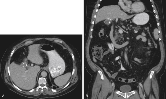
1. What should be included in the differential diagnosis of the imaging finding shown in Figure A? (Choose all that apply.)
2. What serious complication is associated with porcelain gallbladder?
3. What is the most common malignancy of the biliary tract?
D. Extrahepatic cholangiocarcinoma
4. What is the prognosis of gallbladder carcinoma?
B. Mean survival time is about 6 months.
C. The disease is cured by cholecystectomy.
D. Disease progresses to gallbladder perforation.
ANSWERS
CASE 183
Carcinoma of the Gallbladder
1. A, B, C, and D
2. D
3. C
4. B
References
Furlan A, Ferris V, Hossseinzadeh K, et al: Gallbladder carcinoma update: multimodality imaging evaluation, staging, and treatment options. AJR Am J Roentgenol. 2008;191(5):1440-1447.
Cross-Reference
Gastrointestinal Imaging: THE REQUISITES, 3rd ed, p 250.
Comment
Although carcinoma of the gallbladder is uncommon, it is the most common malignant biliary disease. It is generally an adenocarcinoma. Patients present with symptoms that are very similar to pancreatic head lesions: jaundice, weight loss, and abdominal pain. The condition of chronic cholecystitis that leads to cystic duct occlusion and (if it does not result in a septic catastrophe) can eventually result in a calcified gallbladder wall—the porcelain gallbladder (see figures). The risk of gallbladder cancer in this setting is high (about 12%). However, other conditions (if not being direct risk factors) are associated with carcinoma of the gallbladder. About 75% of patients with gallbladder cancer have gallstones, especially a high percentage of cholesterol stones.
Owing to the anatomic position of the gallbladder in or around the porta hepatis, the dissemination of the disease to important contiguous structures makes the outlook for survival bleak. CT is not specific but is suggestive. In some cases, a soft tissue–filled gallbladder, still maintaining its gallbladder configuration (the “jam-packed gallbladder”) may be seen. In many instances, the finding is an amorphous mass in the porta hepatis with bile duct obstruction. The origin may be pancreatic or biliary. Depending on the clinical presentation, severe gallbladder inflammation would also be a consideration.


