CASE 182
History: A 74-year-old woman presents with intermittent abdominal pain following meals.
1. What should be included in the differential diagnosis of the imaging finding shown in the figures? (Choose all that apply.)
E. Nonsteroidal anti-inflammatory drugs
2. What is the most common cause of acute mesenteric ischemia?
A. Superior mesenteric artery thrombosis
B. Superior mesenteric artery embolism
C. Mesenteric venous thrombosis
3. What is the most common CT finding of bowel ischemia?
4. Which of the following statements regarding chronic mesenteric ischemia is true?
A. Chronic mesenteric ischemia is most commonly due to recurrent mesenteric artery emboli.
B. The typical presentation is postprandial crampy abdominal pain.
C. MR angiography is the gold standard imaging modality.
D. Treatment is surgical resection of the ischemic segment of bowel.
ANSWERS
CASE 182
Small Bowel Ischemic Stricture
1. A, B, D, and E
2. B
3. A
4. B
References
Makanjuola D. Computed tomography compared with small bowel enema in clinically equivocal intestinal obstruction. Clin Radiol. 1998;53(3):203-208.
Cross-Reference
Gastrointestinal Imaging: THE REQUISITES, 3rd ed, p 115.
Comment
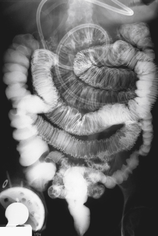
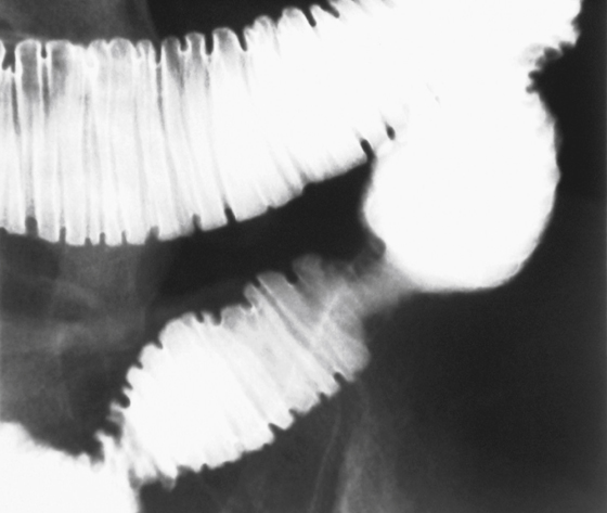
This patient presented with an incomplete small bowel obstruction. A barium small bowel examination showed a focal area of narrowing in the ileum (see figures). The fold pattern is distorted but intact without evidence of mucosal destruction, suggesting this is not a malignant obstruction. The possibility of an inflammatory obstruction was considered. The segment of involved ileum was resected, and pathologic examination of the narrowing showed it to be an area of fibrosis and scarring, resulting from prior ischemic disease. These types of small bowel obstructions are uncommon compared with postoperative adhesions causing small bowel obstruction.
Ischemic small bowel obstruction is seen mostly in elderly patients and results from occlusive or low-flow mesenteric arterial disease. However, focal ischemic and resultant fibrotic strictures of the small bowel have been reported in collagen vascular disease and any condition resulting in a vasculitis, such as rheumatoid arthritis, post-transplant cases and trauma cases without perforation, radiation cases, and some malabsorption processes. Ischemic disease of the colon is more likely to result in a residual stricture (40% of cases). Small bowel obstruction is also a well-known complication of neoplasms, such as primary adenocarcinoma and carcinoids, and inflammatory processes such as Crohn’s disease.


