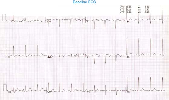
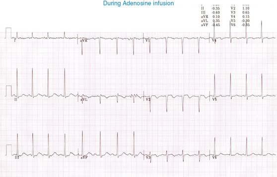
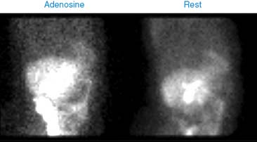
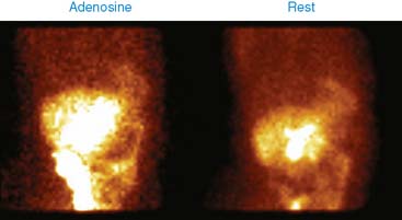
Case 18

Published on 02/03/2015 by admin

Filed under Internal Medicine

Last modified 22/04/2025

Print this page

rate 1 star rate 2 star rate 3 star rate 4 star rate 5 star
Your rating: none, Average: 0 (0 votes)

This article have been viewed 1037 times

Case 18

A 54-year-old woman with inadequately controlled hypertension for the last several years complains of exertional angina at the outpatient office and is referred for pharmacologic stress/rest perfusion imaging. She is on oral atenolol 50 mg per day.

She underwent 2-day rest/stress 99mTc-sestamibi perfusion imaging using 5-minute adenosine infusion as an outpatient study. Her heart rate increased from 83 to 93 beats/min and blood pressure from 185/99 to 179/98 mm Hg. She developed anginal chest pain during adenosine infusion. Her baseline and peak stress ECGs are shown. What is your interpretation?

There is normal sinus rhythm with Q waves in aVL and V2, with T-wave inversion in leads aVL and V2-3. There is left ventricular hypertrophy. There is no ST-segment depression with adenosine infusion.

Perfusion imaging shows a large area of ischemia involving the anterior wall and apex. There is transient hypokinesia involving the anterior wall and apex, which normalizes at rest. LVEF is 51% on the poststress images and 59% on the rest images. This is a high-risk abnormal study.

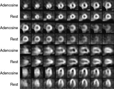
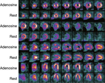
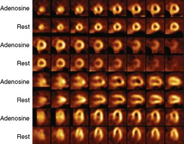
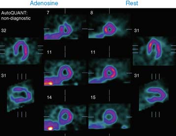
What should the nuclear cardiology laboratory do about this patient?

She is at high risk for developing adverse cardiac events in the short term as well as the long term. Apart from sending the printed test report, to avoid any delays in communication, the interpreting physician should immediately communicate personally by phone call with the referring physician regarding the test result. This was done in this case. Her antianginal medication was increased, and she was brought back to the hospital 1 day later for cardiac catheterization.

She underwent cardiac catheterization, which showed single-vessel coronary artery disease (CAD). There was smooth, 60% narrowing in the distal segment of the left anterior descending (LAD) coronary artery.

However, there was anterolateral wall akinesia that could not be explained by angiographic disease. Although angiographically noncritical, due to her symptoms of angina and evidence of inducible ischemia, intervention was undertaken with percutaneous transluminal coronary angioplasty (PTCA) and a drug-eluting stent.

If there is no prior evidence of ischemia, during catheterization, fractional flow reserve (FFR) can be used to guide intervention in noncritical intermediate coronary stenosis.