CASE 18
1. Which of the following would require coronary catheterization after coronary CT angiography? (Choose all that apply.)
A. 40% stenosis of left anterior descending coronary artery
B. 20% stenosis of left anterior descending coronary artery
C. 60% stenosis of left anterior descending coronary artery
D. 70% stenosis of left anterior descending coronary artery
2. What is the major benefit of coronary CT angiography in the evaluation of a low-risk patient with chest pain?
A. High negative predictive value
B. High positive predictive value
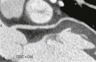
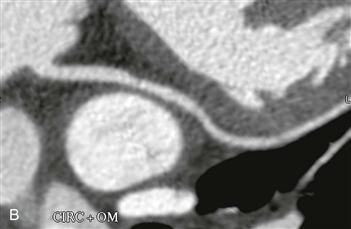
3. What is the finding in the left circumflex coronary artery?
A. Less than 50% narrowing by noncalcified plaque
B. Less than 50% narrowing by calcified plaque
C. Greater than 50% narrowing by noncalcified plaque
D. Greater than 50% narrowing by calcified plaque
4. What is the most appropriate recommendation for this patient?
ANSWERS
Reference
Zimmet JM, Miller JM. Coronary artery CTA: imaging of atherosclerosis in the coronary arteries and reporting of coronary artery CTA findings. Tech Vasc Interv Radiol. 2006;9(4):218–226.
Cross-Reference
Cardiac Imaging: The REQUISITES, ed 3, pp 248–261.
Comment
Imaging
Two multiplanar reformatted images show a short segment stenosis with 20% to 30% diameter reduction in the proximal left circumflex coronary artery. The plaque is eccentric and noncalcified (Figs. A and B).
Diagnosis
The main value of CT angiography is excluding significant coronary artery disease in patients presenting with chest pain who are deemed to have a low or intermediate risk of having had a coronary event. CT angiography is able to exclude disease because of its high negative predictive value in this specific patient population. The degree of vessel stenosis is determined by comparing the luminal diameter at the site of greatest narrowing with the luminal diameter of the most proximal normal segment of coronary artery. A positive CT angiography result is defined as a stenosis with luminal diameter reduction greater than or equal to 50%. Patients with a positive examination must be evaluated further with catheter angiography to confirm and define the abnormality further and potentially to treat the culprit lesion.


