CASE 172
History: A 68-year-old man presents with peritoneal signs, pain, and fever.
1. What should be included in the differential diagnosis of the imaging finding shown in the figures? (Choose all that apply.)
2. Which of the following statements regarding perforated colon carcinoma is false?
A. Perforation is a serious, often fatal complication of colonic carcinoma.
B. Perforation is an uncommon complication of primary colonic carcinoma.
3. What mode of disease spread occurs with the greatest increased incidence in patients with colon cancer presenting with perforation?
4. All of the following are potential causes of a pericolonic fluid collection except:
ANSWERS
CASE 172
Perforated Colon Carcinoma
1. A, B, C, and E
2. D
3. A
4. A
References
Kriwanek S, Armbruster C, Dittrich K, et al: Perforated colorectal cancer. Dis Colon Rectum. 1996;39(12):1409–1414.
Cross-Reference
Gastrointestinal Imaging: THE REQUISITES, 3rd ed, p 302.
Comment
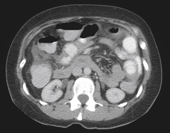
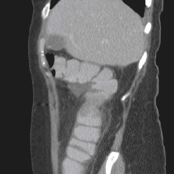
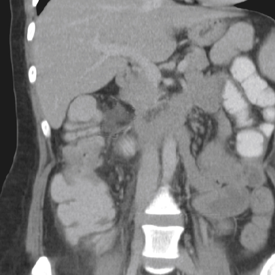
The CT images show inflammatory changes around the colon forming a pericolic abscess (see figures). Although the usual presenting symptoms of obstruction and bleeding (occult or frank bleeding) account for most colonic malignant lesions, perforation is the presentation in a small but significant number of cases (1% to 2%). In most cases, such as in the case presented here, the perforation site is at the site of the tumor. However, perforation proximal to the tumor can be seen in about 30% of these patients. Perforation distal to the tumor is extremely rare. Secondary perforation after radiation treatment has been reported but is also rare.
Perforation linked to colonic carcinoma occurs slightly more frequently on the right side. In such a scenario, the question of a perforated carcinoma is always an issue and must be excluded before considering right-sided diverticulitis. However, when the perforation is left sided and especially in the sigmoid colon, it can mimic diverticulitis almost perfectly.


