CASE 171
History: A 71-year-old woman presents with dysphagia.
1. What should be included in the differential diagnosis of the imaging finding shown in Figure A? (Choose all that apply.)
2. Where in the esophagus are strictures most commonly seen?
3. In what syndrome are cervical esophageal webs associated with iron deficiency anemia?
4. What is the treatment for cervical esophageal webs?
ANSWERS
CASE 171
Esophageal Web
1. A, B, and E
2. A
3. B
4. B
References
Luedtke P, Levine MS, Rubesin SE, et al: Radiologic diagnosis of benign esophageal strictures: a pattern approach. Radiographics. 2003;23(4):897–909.
Cross-Reference
Gastrointestinal Imaging: THE REQUISITES, 3rd ed, p 32.
Comment
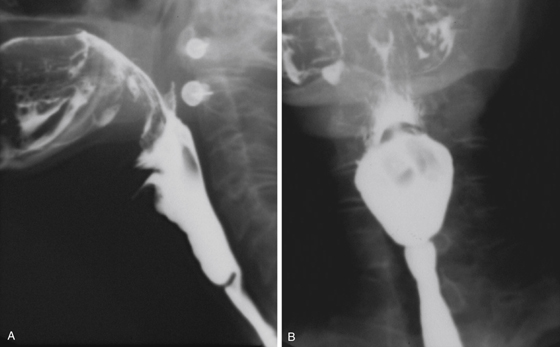
Images from a barium esophagogram show a well-defined web or ring stricture in the proximal esophagus (see figures). These proximal webs are common. If the web is small or noncircumferential, the patient may be asymptomatic. However, if the lumen is sufficiently compromised, such that a 13-mm barium tablet would be held up by the web, the patient is likely to complain of dysphagia. These webs are seen accompanied by iron deficiency anemia in Plummer-Vinson syndrome (also known as Paterson-Kelly syndrome in the United Kingdom). The condition is sometimes referred to as sideropenic dysphagia.
The exact cause of this condition is unclear. Some authors doubt the validity of the association or the syndrome altogether. Plummer-Vinson syndrome has been speculated to be an autoimmune phenomenon, but no conclusive evidence has been offered to prove this hypothesis. The condition was first described and named in the early 20th century, when the incidence was thought to be common. It is now seen rarely, possibly because fewer barium esophagograms are being done, and endoscopy may not see the web or may push by it (and effect a cure), or, alternatively, Plummer-Vinson syndrome may be a nonentity.
Authors who have described the condition have mostly described an esophageal web seen in the cervical esophagus during a barium esophagogram. It has been said to be found predominantly in white women with a predisposition for increased risk of squamous cell carcinoma of the esophagus. However, this risk factor is also disputed. For the radiologist, it is important to rule out strictures related to other conditions, such as skin lesions, epidermolysis bullosa, and pemphigus, and strictures related to gastroesophageal reflux.


