CASE 170
History: A 40-year-old woman presents with right upper quadrant pain and weight loss.
1. What should be included in the differential diagnosis of the imaging finding shown in the figure? (Choose all that apply.)
2. What is the most common tumor of the liver of vascular origin?
3. Which of the following describes the prognosis of hepatic epithelioid hemangioendothelioma (EHE)?
A. Intermediate between benign and malignant
B. Benign until transformation to hepatoma
4. What is the most common site of EHE?
ANSWER
CASE 170
Epithelioid Hemangioendothelioma
1. A, C, and E
2. A
3. A
4. A
References
Lyburn ID, Torreggiani WC, Harris AC, et al: Hepatic epithelioid hemangioendothelioma: sonographic, CT, and MR imaging appearances. AJR Am J Roentgenol. 2003;180(5):1359–1364.
Cross-Reference
Gastrointestinal Imaging: THE REQUISITES, 3rd ed, p 205.
Comment
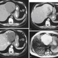
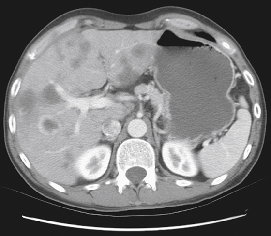
The hepatic lesions are of soft tissue density, and some have a well-defined “halo” around them (see figure). This appearance should not dissuade the radiologist from considering metastatic disease as the first thing to be excluded. However, no primary disease was present in this patient, and biopsy of the liver lesions revealed the multinodular lesions of the liver to be EHE. This is a rare condition mostly seen in soft tissues and bone. Involvement of the liver resembles metastatic disease. Although metastatic lesions can have an enhancing halo, EHE seems to have a more prominent halo sign; however, this does not apply in all hepatic lesions as seen on the transaxial image of the liver.
In a setting in which a primary lesion is not evident, and well-defined peripheral enhancement that is brighter than usual is present (and is not the “puddling” effect seen with benign hemangiomas), EHE is a consideration. The tumor is an unusual vascular lesion characterized by epithelioid endothelial cells. It has been described in adults of all ages. The tumor has the histologic features of a low-grade malignancy. In 2002, the World Health Organization defined “hemangioendothelioma” as a definitive, locally aggressive malignant tumor that rarely metastasizes.
In the liver, EHE is seen as multiple, indolent, slow-growing progressive lesions, which on CT may show a bright, well-defined halo effect. It is extremely rare to see spread of the tumor beyond the liver.