CASE 17


1. What should be included in the differential diagnosis? (Choose all that apply.)
A. Artifactual narrowing of coronary artery
2. What degree of coronary arterial narrowing is considered a positive CT angiography result?
A. 0%
B. 15%
C. 35%
D. 50%
3. What is the finding in the right coronary artery?
A. Normal
D. Dissection
4. What is the best next step for management of this patient with a 50% narrowing?
ANSWERS
Reference
Townsend JC, Gregg D IV. Cardiac computed tomography and magnetic resonance imaging: the clinical use from a cardiologist’s perspective. J Thorac Imaging. 2010;25(3):194–203.
Cross-Reference
Cardiac Imaging: The REQUISITES, ed 3, pp 248–261.
Comment
Imaging
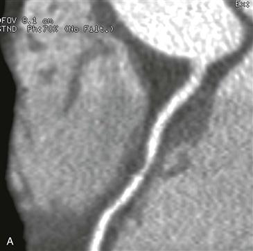
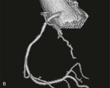
Curved multiplanar reformat and volume rendered images show a focal stenosis with diameter reduction of approximately 50% in the proximal right coronary artery (Figs. A and B). The narrowing consists of noncalcified plaque without calcification.
Further Evaluation
The clinical utility of CT angiography of the coronary arteries is highly dependent on the patient population studied. The diagnostic value of CT angiography is greatest in patients with a low pretest probability of disease and is lowest in patients with a high pretest probability of disease. In other words, CT angiography has a high negative predictive value for excluding disease in patients with a low or intermediate pretest probability of coronary artery disease. Patients with a negative test do not need further evaluation for coronary disease. In this case, the test is positive (≥50% narrowing) and the patient needs a coronary angiogram to confirm the presence of significant disease. Coronary catheterization confirmed right coronary artery stenosis with a 60% diameter reduction, and this was successfully treated with a stent.
Patient Selection
The ideal patient to undergo CT angiography of the coronary arteries is a patient with a low or intermediate pretest probability of coronary artery disease. CT angiography is not indicated in patients with typical findings of coronary artery disease or in patients with a high pretest likelihood of disease (i.e., ECG changes suggestive of ischemia or laboratory evidence of myocardial injury). CT angiography of the coronary arteries is also not indicated in patients suspected to have another disease (i.e., pneumonia, pneumothorax, or pulmonary embolism).


