CASE 165
History: A 65-year-old woman presents with abdominal distention.
1. What should be included in the differential diagnosis of the imaging finding shown in Figure A? (Choose all that apply.)
2. What process within the liver gives rise to the appearance of the nutmeg liver?
A. Chronic systemic venous congestion
B. Chronic portal venous congestion
C. Chronic arterial obstruction
D. Chronic biliary obstruction
3. What is the most common disease resulting in chronic systemic venous congestion?
C. Systemic arterial hypertension
D. Pulmonary venous congestion
4. Which of the following features suggests Budd-Chiari syndrome instead of systemic venous congestion as the cause of a diffuse mottled nutmeg liver appearance?
D. Distention of the inferior vena cava
ANSWERS
CASE 165
Nutmeg Liver
1. A, B, C, and E
2. A
3. B
4. C
References
Brancatelli G, Vilgrain V, Federle MP, et al: Budd-Chiari syndrome: spectrum of imaging findings. AJR Am J Roentgenol. 2007;188(2):W168–W176.
Liu WL, Lai CC. Images in emergency medicine: nutmeg liver. Emerg Med J. 2011.28.
Cross-Reference
Gastrointestinal Imaging: THE REQUISITES, 3rd ed, p 183.
Comment
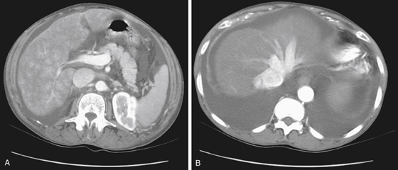
Right-sided heart failure is almost always the basis of a diffuse mottled-appearing, enlarged liver, with distended hepatic veins (see figures). It has been referred to by pathologists as nutmeg liver for more than 100 years and is a very common finding at autopsy. Any chronic preterminal event may result in right-sided heart failure—hence the high incidence seen at autopsy. Nutmeg is a spice, the germ taken from the seed of a tree found in the tropics. The name “nutmeg liver” was used by pathologists to describe sections taken from a chronically passive congested liver, usually caused by right-sided heart failure. The congestion and pigmentation around the central veins of the liver lobules gave it a “nutmeg” appearance to the eyes of early pathologists, who were inclined to give food-related names to disease entities in the early days of histopathology (i.e., “bread and butter pericarditis,” “anchovy sauce” liver lesions).
The nutmeg liver would have remained an interesting sidelight of histopathology if not for the advent of CT imaging. When evaluating patients with congestive heart failure, especially right-sided failure, radiologists began to observe that the liver took on a diffuse mottled appearance almost identical to that described by the pathologists. Distention of the hepatic veins and degrees of hepatomegaly were also seen.
The potential problem of the nutmeg liver is that it could conceivably mask some other hepatic disease. The term “nutmeg liver” is now part of the lexicon of radiology and hepatic imaging.


