CASE 161
History: A 61-year-old woman presents for evaluation of fainting episodes.
1. What should be included in the differential diagnosis of the imaging finding shown in the figures? (Choose all that apply.)
A. Metastases from a neuroendocrine tumor
B. Metastases from a mucinous carcinoma
C. Multifocal hepatocellular carcinoma
2. Which of the following is the most common pancreatic islet cell tumor?
D. Nonfunctioning islet cell tumor
3. What is the most common malignancy that results in hypervascular liver metastases?
D. Pancreatic islet cell tumor
4. If this patient exhibits palpitations, sweating, and headache, what clinical diagnosis should be suspected?
ANSWERS
CASE 161
Islet Cell Tumor of the Pancreas
1. A, B, C, and D
2. D
3. A
4. B
References
Horton KM, Hruban RH, Yeo C, et al: Multi-detector row CT of pancreatic islet cell tumors. Radiographics. 2006;26(2):453–464.
Cross-Reference
Gastrointestinal Imaging: THE REQUISITES, 3rd ed, p 164.
Comment
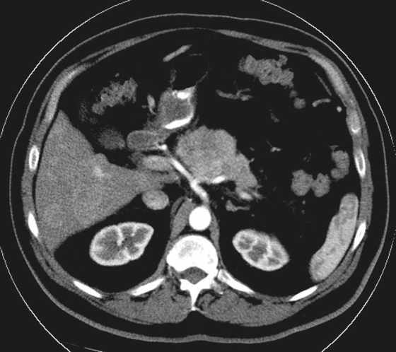
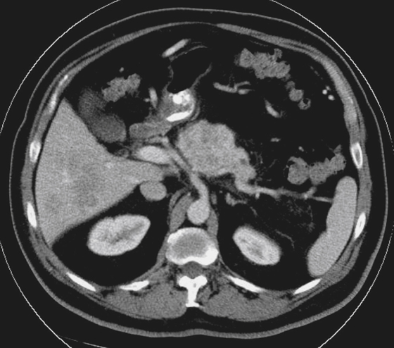
About 50% of all islet cell tumors of the pancreas are nonfunctioning islet cell tumors. Because they are relatively asymptomatic (they do not produce and secrete endocrine products), they can grow to large size, such as in this case (see figures). The biliary system is not invaded, and there is no biliary dilation within the liver. Because these tumors grow slowly and quietly over a long period, they are quite large when discovered and may be malignant. Studies have shown that almost 50% of islet cell tumors of the pancreas are malignant at the time of discovery, mostly by contiguous spread or from metastatic disease to the liver.
Most tumors are large enough to have necrotic centers at the time of discovery. In addition, nonfunctioning islet cell tumors may have a cystic component, which can confuse and delay diagnosis. Nonfunctioning islet cell tumors can occur anywhere in the pancreas, with the most popular sites being the head and tail. They can reach sizes of 8 to 20 cm.
Even with the high potential of malignancy, the prognosis remains good, which is not the case for ductal adenocarcinomas of the pancreas. Of all islet cell tumors, functioning or nonfunctioning, 20% to 25% have some calcifications within them, whereas only 1% to 2% of ductal carcinomas show any sign of calcification. The treatment is surgical.


