Case 16

Published on 02/03/2015 by admin

Filed under Internal Medicine

Last modified 22/04/2025

Print this page

rate 1 star rate 2 star rate 3 star rate 4 star rate 5 star
Your rating: none, Average: 0 (0 votes)

This article have been viewed 1011 times

Case 16

A 76-year-old male with known coronary artery disease (CAD) and multiple myocardial infarctions (MIs) and congestive heart failure (CHF) in the past was admitted to the hospital because of chest pain and worsening heart failure. He underwent cardiac catheterization, which showed 100% occlusion of the right coronary artery (RCA) and left circumflex (LCX) and 80% narrowing of the left anterior descending (LAD). Left ventriculogram showed severely impaired left ventricular ejection fraction (LVEF) of 15%.

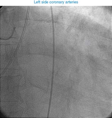
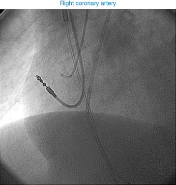
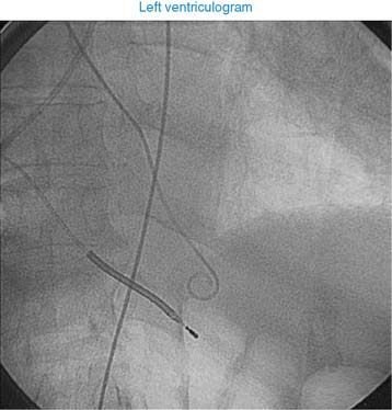
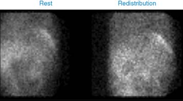
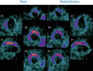
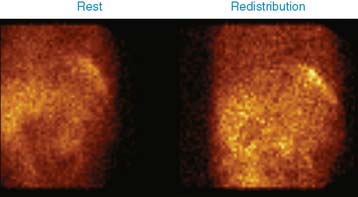
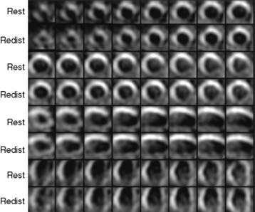
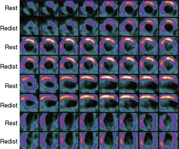
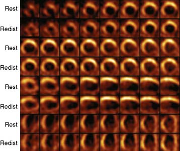
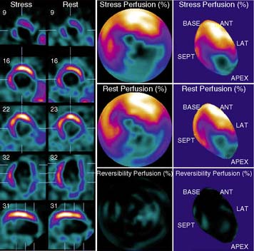
He was referred for a viability study to decide the further course of management and possible revascularization. A dose of 3.2 mCi of thallium-201 was injected at rest, followed by gated SPECT imaging 15 minutes and 4 hours later (Videos 4 and 5 and Figures 1 and 2). What is your interpretation of this study?

The left ventricle (LV) is massively enlarged. There is a very large, dense area of perfusion abnormality involving the inferior and lateral walls and apex, which does not change on delayed images. The tracer uptake in septum is decreased, which does not change on delayed imaging. Tracer uptake is normal in the anterior wall. The LV is severely hypokinetic and LVEF is depressed at 7%. This study indicated dense scar involving the inferior and lateral walls and apex with no evidence of viability. The septum shows an admixture of scar and viable myocardium, and the anterior wall is normally perfused.

This study reveals a very large, dense scar with no evidence of viability involving the inferior and lateral walls and apex, which correspond to completely occluded RCA and LCX. The septum and anterior wall show viability. These walls are perfused by a severely narrowed LAD. Revascularization of the LAD may improve the perfusion and possibly the function of the septum and anterior wall. However, the revascularization of the LAD in this case would be a very high-risk procedure, and global LVEF may not show any improvement, given the presence of a very large, dense scar involving the inferior and lateral walls.

The patient opted to be treated medically without any revascularization. His heart failure continued to worsen, and he died of refractory heart failure.