History: A 57-year-old male alcoholic presents with a 6-month history of retrosternal dysphagia and 10-kg weight loss.
1. What should be included in the differential diagnosis of the imaging finding shown in the figure? (Choose all that apply.)
C. Downhill esophageal varices
2. What part of the bowel wall present in other segments of the GI tract is not found in the esophagus?
3. Why do “jump” metastases occur in esophageal carcinoma?
A. Portosystemic venous collaterals
B. Network of lymphatic channels
C. Absence of esophageal serosa
D. Metachronous tumor deposits
4. What disease is the world’s most common cause of portal hypertension and varices?
ANSWERS
CASE 158
Esophageal Varicoid Cancer
1. A, B, C, and E
2. D
3. B
4. C
References
Iyer R, Dubrow R. Imaging upper gastrointestinal malignancy. Semin Roentgenol. 2006;41(2):105–112.
Levine MS. Esophageal cancer: radiologic diagnosis. Radiol Clin North Am. 1997;35(2):265–279.
Cross-Reference
Gastrointestinal Imaging: THE REQUISITES, 3rd ed, p 32.
Comment
Esophageal carcinoma can appear in many different forms. The most common form is a stricture, usually irregular in nature. It also may be an eccentric mass or a polypoid mass within the lumen. It may ulcerate, with resultant bleeding. The anatomy of the esophagus is different from the anatomy of the remainder of the GI tract, which results in the rapid spread of the neoplasm and the resultant poor prognosis.
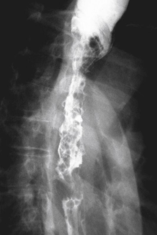
In contrast to the rest of the GI tract, there is no serosal layer in the esophagus, and neoplasms invade the adjacent structures directly, resulting in a high rate of morbidity. Also, the lymphatic drainage of the esophagus is complex. There is an extensive network of lymphatic channels in all layers of the esophagus. This characteristic results in metastases spreading circumferentially and to adjacent lymph node groups in the mediastinum. The tumor may “jump” or disseminate throughout the length of the esophagus via these channels. There may be intervening normal mucosa between these areas of tumor. This pattern of spread is believed to result in the varicoid appearance of esophageal carcinoma seen in this patient (see the figure). Despite the history of alcoholism in this patient, varicoid carcinoma of the esophagus can be distinguished by the fact that the pattern does not change whether the patient is upright or recumbent, does not change with Valsalva respiration, is not pliable, and carries no peristaltic waves.
Varicoid carcinoma is an unusual variant of esophageal carcinoma. The tumor spreads submucosally down the length of the esophagus, producing thickened folds. The appearance closely mimics esophageal varices—hence the name. Because of this pattern of spread, dysphagia is usually a late symptom, and the disease has usually spread extensively before being diagnosed. A patient with this form of esophageal neoplasm has a poor prognosis.


