CASE 156
History: A 56-year-old woman presents with abdominal distention.
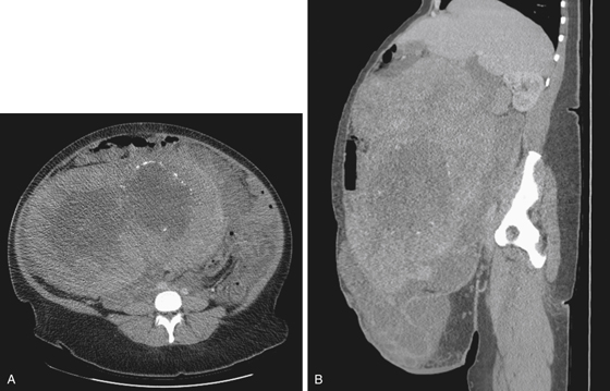
1. What should be included in the differential diagnosis of the site of origin of the imaging finding shown in Figure A? (Choose all that apply.)
2. What is the most common gynecologic tumor in women older than 30 years?
3. What is the most common mode of presentation of uterine fibroids?
4. What imaging modality would you recommend to visualize uterine fibroids best when planning for surgery or intervention?
ANSWERS
CASE 156
Huge Myometrial Leiomyomas
1. A, B, D, and E
2. C
3. A
4. D
References
Fielding JR. MR imaging of the female pelvis. Radiol Clin North Am. 2003;41(1):179–192.
Cross-Reference
Gastrointestinal Imaging: THE REQUISITES, 3rd ed, p 308.
Comment
Uterine fibroids do not usually present a problem for either patients or radiologists. They are commonly a secondary finding noted at the time of examination (especially in CT). This patient’s huge myometrial leiomyoma is unusual, and malignancy was suspected (see figures). A 30-lb fibroid and adhesions and fluid were found at surgery, and pathologic examination showed no evidence of malignancy. There is a small malignant risk in uterine fibroids usually reported as about 1%. Most are asymptomatic. Some are implicated in infertility states.
Common treatments include selective resection or total hysterectomy, if deemed necessary. An estimated 600,000 hysterectomies are performed annually in the United States, and approximately one-third are for uterine leiomyomas (fibroid). In recent years, treatment by uterine artery embolization has proved helpful in the treatment of symptomatic lesions.
CT and ultrasound are often used in the evaluation of this condition. However, MRI may also be very helpful. Fibroids tend to enhance on the margins when gadolinium enhancement is used. The rim may be seen in unenhanced cases with T2 imaging. The tissue signal is usually lower than the surrounding tissue on both T1 and T2 imaging. Small linear arrangements of calcification can be seen in this lesion, which might suggest benignity but cannot rule out malignancy. Alternative diagnostic considerations in this patient would be ovarian mass, tumor originating from ovary or uterus, uterine leiomyosarcoma, and adenomyosis of the uterus.


