CASE 155
1. What should be included in the differential diagnosis? (Choose all that apply.)
A. Transposition of the great arteries
2. Based on the radiograph, what is the most likely diagnosis?
A. Transposition of the great arteries
3. What is the most common right aortic arch branching associated with this anomaly?
B. Aberrant innominate (brachiocephalic) artery
C. Aberrant left subclavian artery
D. Isolated left subclavian artery
4. Which of the following CT techniques would reduce radiation dose?
ANSWERS
Reference
Johnson TR. Conotruncal cardiac defects: a clinical imaging perspective. Pediatr Cardiol. 2010;31:430–437.
Cross-Reference
Cardiac Imaging: The REQUISITES, ed 3, pp 324–327.
Comment
Anatomy and Clinical Features
Truncus arteriosus is a cyanotic admixture lesion in which the pulmonary artery, aorta, and coronary arteries arise from a common trunk. There may be a main pulmonary artery, but in some patients the right and left pulmonary arteries arise separately from the common trunk. A ventricular septal defect is present in this anomaly. The truncal valve usually is tricuspid, but it can have four or more leaflets. Approximately 30% to 35% of patients have a right aortic arch, which most commonly has mirror-image branching. Truncus arteriosus is classified into different types according to the pulmonary artery anatomy. Treatment is surgical correction within the first year of life.
Associations
There is an association with DiGeorge syndrome and interrupted aortic arch. Other abnormalities may involve the coronary arteries, mitral valve, and pulmonary venous connections.
Imaging
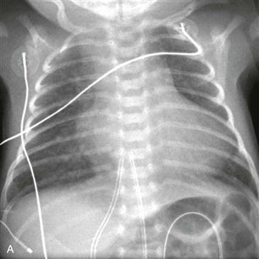
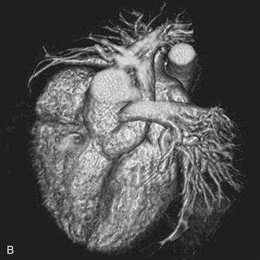
Radiographs typically show shunt vascularity, cardiomegaly, and frequently a right-sided aortic arch (Fig. A). Echocardiography is generally adequate for diagnosis and preoperative planning in most patients. CT (Fig. B) or MRI may be performed in difficult cases to delineate the anatomy of the branch pulmonary arteries or aorticopulmonary collaterals and in patients with complex aortic arch anomalies.
Role of Imaging after Corrective Surgery
The major role of CT and MRI in truncus arteriosus is in detecting complications after corrective surgery. Important complications include right or left ventricular dysfunction, pulmonary homograft stenosis, pulmonic insufficiency, branch pulmonary artery stenosis, and neoaortic valve dysfunction.


