CASE 152
1. What should be included in the differential diagnosis based on the pattern of chamber opacification? (Choose all that apply.)
C. Partial anomalous pulmonary venous connection
2. Which cardiac chamber should opacify first after contrast agent injection into the umbilical vein catheter?
A. Left atrium
C. Right atrium
3. What is the purpose of the foramen ovale?
A. To allow right-to-left shunting in fetal life
B. To allow left-to-right shunting in fetal life
C. To allow right-to-left shunting in postnatal life
D. To allow left-to-right shunting in postnatal life
4. If the foramen ovale remains patent, what is the most significant complication?
A. Cyanosis
ANSWERS
References
Berko NS, Haramati LB. Simple cardiac shunts in adults. Semin Roentgenol. 2012;47(3):277–288.
Kim YJ, Hur J, Shim CY, et al. Patent foramen ovale: diagnosis with multidetector CT—comparison with transesophageal echocardiography. Radiology. 2009;250(1):61–67.
Kutty S, Sengupta PP, Khandheria BK. Patent foramen ovale: the known and the to be known. J Am Coll Cardiol. 2012;59(19):1665–1671.
Cross-Reference
Cardiac Imaging: The REQUISITES, ed 3, p 335.
Comment
Clinical Features
In fetal life, the patent foramen ovale provides a channel for umbilical vein blood to bypass the lungs and enter the systemic circulation directly. The foramen is covered by a flap that should close after birth. However, the flap does not close in a substantial minority of individuals, and approximately 25% of adults have a patent foramen ovale. The most important complication of this condition is paradoxical embolism, which can result in ischemia or infarction in one or more organs, including the brain. Some patients are treated with a transcatheter septal occluder device.
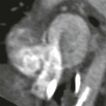
Imaging Findings
Echocardiography and occasionally CT (Figure) and MRI can demonstrate blood flowing across the foramen ovale. The direction of the shunt can be from left to right in a substantial percentage of people or may reverse (as in the case shown) when right atrial pressure exceeds left atrial pressure as in coughing or with the Valsalva maneuver. The patent foramen ovale typically has a small channel-like configuration on CT or MRI. To make a confident diagnosis on CT, both the channel-like configuration in the interatrial septum and a jet of contrast medium passing between the atria must be observed. The standard reference, transesophageal echocardiography with agitated saline, detects a patent foramen ovale by momentarily seeing microbubbles passing across the interatrial septum into the left atrium during provocative maneuvers. Many clinicians believe that echocardiography may miss a substantial percentage of cases of patent foramen ovale, especially in patients who cannot cooperate with Valsalva instructions.


