CASE 150
History: A patient presents with chest pain.
1. What should be included in the differential diagnosis? (Choose all that apply.)
2. What is the most common cause of a discrete sinus of Valsalva aneurysm?
B. Syphilis
C. Weakness at the junction of the aorta and valve
3. What valvular lesion is most commonly associated with a sinus of Valsalva aneurysm?
4. Which type of ventricular septal defect (VSD) is associated with a discrete sinus of Valsalva aneurysm?
A. Muscular
D. Supracristal
ANSWERS
References
Hoey ET, Kanagasingam A, Sivananthan MU. Sinus of Valsalva aneurysms: assessment with cardiovascular MRI. AJR Am J Roentgenol. 2010;194(6):W495–W504.
Moustafa S, Mookadam F, Cooper L, et al. Sinus of Valsalva aneurysms—47 years of a single center experience and systematic overview of published reports. Am J Cardiol. 2007;99(8):1159–1164.
Cross-Reference
Cardiac Imaging: The REQUISITES, ed 3, pp 389–392.
Comment
Etiology and Pathology
Discrete sinus of Valsalva aneurysms usually result from congenital weakness at the junction of the aortic media and the annulus fibrosus of the valve. They most frequently arise from the right coronary or noncoronary sinuses. Aneurysms of the right coronary sinus rupture into the right atrium or right ventricle, and aneurysms of the noncoronary sinus rupture into the right atrium. Sinus of Valsalva aneurysms are associated with aortic regurgitation. They can occur in patients who have a supracristal VSD.
Imaging
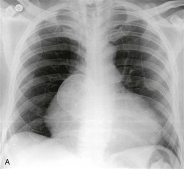
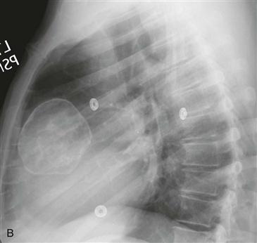
Chest radiography may be normal or may show a mass arising from the right side of the mediastinum (Figs. A and B). On cross-sectional imaging examinations, a discrete sinus of Valsalva aneurysm manifests as a focal dilation of one sinus of Valsalva, in contrast to the generalized aortic root dilation seen in annuloaortic ectasia. Calcification can be seen in these lesions (Figs. A and B).


