CHAPTER 15. NASOGASTRIC TUBE PLACEMENT
Indications124
Contraindications124
Equipment124
Practical procedure124
Post-procedure investigations127
Complications127
The modern nasogastric (NG) tube is a 1921 modification by John Alfred Ryle (1889–1950). He covered the tip of the NG tube with rubber to prevent injury to the gastric mucosa, unlike Previous tubes which had a metal bulb at their tip. However, the earliest recording of using a tube for enteral nutrition was that of the Seville-born Arab surgeon Ibn Zuhr (also known as Avenzoar, 1091–1161) who fed a patient with an oesophageal stricture via a silver tube. Incidentally Ibn Zuhr is also credited with the first correct description of performing a tracheotomy for suffocating patients.
INTRODUCTION
Nasogastric tube placement is a simple procedure, but can be unpleasant for the conscious patient. This is commonly performed by nursing staff; however, junior doctors would be expected to place them if these initial attempts are unsuccessful. Small-diameter (8–12 Fr) tubes are frequently used for patents who require enteral feeding. Larger tubes (14 Fr or larger) are used to administer medications, provide gastric decompression or allow continuous aspiration of retained gastric contents. These larger tubes are acceptable for feeding over a short period, usually less than 1 week. Small-bore NG tubes cause less trauma to the nasal mucosa both during insertion and while in situ, and are better tolerated. Placement errors lead to potentially major complications, most commonly when mistakenly placed in the respiratory tract. Failure to observe pathological states that constitute a contraindication to NG tube placement can result in passage of the tube into the brain (base of skull fracture) or peritoneum (upper GI perforation).
INDICATIONS
WIDE-BORE NASOGASTRIC TUBES
• Bowel obstruction.
• Gastric outlet obstruction.
FINE-BORE NASOGASTRIC TUBES
• Enteric feeding.
CONTRAINDICATIONS
• Coagulopathy (a relative contraindication).
• Oesophageal varices.
• Maxillofacial and oropharyngeal trauma or surgery.
• Skull fractures.
• Unstable cervical spine injury.
• Laryngectomy.
• Compromised airway.
EQUIPMENT
• Sterile gloves.
• Kidney bowl.
• Gauze.
• Lubricant jelly.
• NG tube.
• 10 mL syringe.
• Litmus paper.
• Medical adhesive tape.
• Drainage bag.
• Gloves.
• Glass of water with straw.
PRACTICAL PROCEDURE
• Explain the procedure to the patient and obtain consent.
• Wash your hands and wear sterile gloves.
• The patient should be in an upright position, the head supported with pillows if needed to ensure that it is not tilting backwards.
• Check the patency of the nostrils; ask the patient to blow their nose if needed.
• Lubricate the NG tube tip and the first 10 cm.
• Slowly insert the NG tube into a nostril using a rotating motion and advance it horizontally along the base of the nasal cavity until it reaches the posterior pharynx (and hence usually induces the gag reflex).
 Tip Box
When the NG tube is in the pharynx, rotate the tube 180 degrees to encourage passage of the tube into the oesophagus rather than the trachea. This is especially useful if the tube has been refrigerated, making it more rigid.
 Tip Box
If you are having trouble passing the NG tube, try asking the patient to put their chin to their chest and insert the tube as above until it reaches the posterior pharynx.
 Tip Box
If you are experiencing trouble passing the tube into the oesophagus because it is too soft and it keeps folding on itself in the back of the throat, try putting the tube in the fridge for 20 minutes for it to harden, or consider a larger tube.
• Ask the patient to swallow water repeatedly through a straw whilst advancing the NG tube during the swallow. In this manner the patient effectively swallows the tube into the oesophagus.
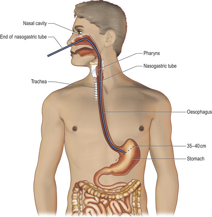
• Advance the NG tube down the oesophagus and into the stomach (Fig. 15.1). The NG tube is graduated every 10 cm (usual mouth to stomach distance is 35–40 cm). Hence insert the NG tube to the 40 cm mark.
• Ensure the NG tube is sitting in the stomach:
— Gently aspirate stomach contents with a syringe and test with litmus paper (if in the stomach the pH should be less than 4). Remember, patients on proton pump inhibitors, antacids and H2 antagonists may have a high gastric pH and hence aspiration tests may not be accurate. X-ray confirmation may have to be used in these patients.
— Small-bore NG tubes commonly do not allow aspiration of stomach contents and require chest X-ray confirmation of position.
— If you are unable to get adequate stomach contents to test for pH, the patient will need to have a chest X-ray to check the position of the tube. The tip of the tube must be seen to be below the diaphragm.
• Attach the drainage bag.
• Secure the NG tube around the patient’s nose with micropore tape taking care not to exert too much pressure on the nares adjacent to the NG tube.
POST-PROCEDURE INVESTIGATIONS
• If you are in any way unsure about the position of the NG tube, obtain a chest X-ray. The NG tube tip has a radio opaque line at the end and can therefore be seen. However, this X-ray confirmation is valid only at the time of the X-ray.
• The NG tube should not be used until positive evidence of its placement has been obtained (pH or chest X-ray).
• Syringing air down the NG tube whist auscultating the epigastrium is an unreliable method of checking tube position due to transmitted lung sounds.
COMPLICATIONS
• Failure to pass the NG tube.
• Epistaxis.
• NG tube passed into trachea.
• Oesophageal, pharyngeal or gastric perforation.
• NG tube in duodenum – should this be confirmed on chest X-ray pull the tube back approximately 5 cm.
• Oesophagitis and stricture formation secondary to oesophageal inflammation.
• Retropharyngeal or nasopharyngeal necrosis.
• Sinusitis from NG tube in situ for a prolonged period of time.


