CHAPTER 15 Electroacupuncture Analgesia
INTRODUCTION
Electroacupuncture is not unanimously defined. Professor Ji-sheng Han of Beijing University states that when electrodes are placed on the surface of the skin, and when “the point of stimulation is selected according to traditional acupuncture, the process is usually called electroacupuncture (EA).” The same process, if the points used are not traditional acupoints, is regarded as transcutaneous electrical nerve stimulation (TENS).1
Neurophysiologically, there is no difference between the two concepts of EA and TENS. The same author also indicated that, “they operate through very similar, if not identical, mechanisms.”1 Different acupoints may have a different anatomic configuration (see Chapter 1), but sensory nerve fibers are the universal component of any acupoint. Because these fibers are distributed all over the body except nails and hair (which is why we can cut them without pain), acupoints can be found anywhere on the body.
If surface electrodes are placed on a sensitized (tender) point, whether it is a traditional acupoint or not, peripheral stimulation will be provided to the spinal cord through the sensitized nerve endings, which helps to desensitize and calm down the irritated nerve. If the surface electrodes are placed on nonsensitive points, the electrophysiologic process resulting from this peripheral stimulation is exactly the same as with the stimulation of tender acupoints, but with less therapeutic results in terms of desensitizing the painful sensory nerves. As we have already mentioned, the number of traditional and extra-meridian “new” acupoints has reached more than 2500 (see Chapter 1), which means that documented acupoints can be found almost everywhere in the body.
Another method also described as electroacupuncture is the use of needles as electrodes, inserted into the tissue, which is called percutaneous electrical nerve stimulation (PENS). Physiologically PENS is different from both EA and TENS because with PENS needles are used to create both tissue lesion and electrical stimulation. Even though EA, TENS, and PENS all stimulate the release of the natural neurochemicals, including endorphins, produced by the central nervous system, PENS with its induced lesions involves other healing mechanisms that are similar to manual needling (see Chapter 3).
Electrotherapy was revived in modern times after Drs. Ronald Melzack and Patrick Wall proposed their Gate Control theory of pain in 1965 (see Chapter 3). This theory stated that the input of pain signals from the fine nerve fibers (such as C fibers or A-δ fibers) is controlled and modified in the spinal cord by the signals from the large nerve fibers (such as A-β fibers) before the pain signals reach the brain. The spinal cord functions like a gate in that it can be open or closed to the incoming pain signals. For example, if a person hits his finger with a hammer, the C nerve fibers in the finger start to fire pain signals to the brain through the spinal cord, so the brain perceives the finger pain. Then the person might rub or scratch the painful finger. By doing this, the large, low-threshold A-β fibers are activated. Their signals travel faster than those of the fine C nerve fibers, and activate the spinal cord to block the pain signals of the C nerve fibers. This is a simplified picture of the Gate Control theory. A recent study using the techniques of molecular biology has supported the concept that endorphins in the spinal cord exert a strong inhibitory effect on incoming pain signals.2
FREQUENCY-DEPENDENT RELEASE OF ENDORPHINS BY PERIPHERAL ELECTRICAL STIMULATION AND THE ANTIENDORPHIN FEEDBACK INTERACTION
The stimulation of EA and TENS is transmitted by A-β and A-δ nerve fibers. Since the parameters of the type of stimulation used with EA and TENS can be reliably and precisely calibrated, it is possible to identify the physiological effects of peripheral electrical stimulation at different frequencies. This frequency-dependent nature of endorphin release has been confirmed in both laboratory animals and human subjects.3
It has been shown that if the stimulation alternates between 2 and 100 Hz, the full release of all four endorphins is achieved, which induces synergistic analgesic effects.4
In addition to opioid peptides, other neurochemical analgesic factors are also triggered by peripheral electrical stimulation. Midbrain monoamines such as serotonin and norepinephrine have been confirmed to play a role in EA analgesia.5,6 Recently it was discovered that brain-derived neurotrophic factor (BDNF) is released by 100 Hz stimulation in bursts, but not by constant 100 Hz stimulation.7 BDNF has been shown to reverse the dying of neurons in animals.8
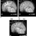
Part of the brain (ventral periaqueductal gray [vPAG]) reacts to both low frequency (2 Hz) and high frequency (100 Hz), but other parts react to only one frequency: the arcuate nucleus of the hypothalamus (ARH) to low frequency and the parabrachial nucleus (PBN) to high frequency.
The body’s wisdom in ensuring the survival mechanism is always ahead of human intelligence. The release of natural endorphins to reduce the body’s pain and stress is a natural physiologic process, but we try to use the electrical stimulation to change this physiological process into a pharmacologic process. Our body has a built-in mechanism to prevent overuse of its natural narcotics (endorphins): the release of antiendorphin peptides through negative feedback. Data from animals show that the analgesic effect of endorphins declines when EA stimulation is prolonged for more than 3 hours due to the release of antiendorphin antagonists (antiopioid peptides).9
Low-frequency stimulation (2 to 15 Hz), triggers the release of endomorphin, enkephalin, and most β-endorphins, but these frequencies also cause the release of their antagonist peptides, substance P (SP), angiotensin II (AII), and cholesystokinin octapeptide (CCK-8). High-frequency (100 Hz) stimulation significantly increases the release of antagonists AII and CCK-8.1
All the laboratory and clinical experimental data indicate that to elicit the maximal release of central opioid peptides (endorphins), electrical stimulation should alternate between 2 Hz and 100 Hz to achieve the synergistic effect of TENS and EA analgesia.1
TECHNICAL PARAMETERS OF TENS AND EA
The purpose of electrical analgesia is to electrically depolarize the nerve endings to produce nerve impulses to the brain through the spinal cord. With a modern EA or TENS apparatus the stimulation parameters can be very easily regulated. The practitioner needs to set up (1) frequencies, (2) modes (constant, burst, or alternative, if they are available), and (3) pulse width. All TENS apparatuses have the option of using square wave or biphasic current (Box 15-1).
Box 15-1 Parameters of Electrical Stimulation Using TENS or EA
Modified from White A: Electroacupuncture and acupuncture analgesia. In Filshie J, White A, editors: Medical acupuncture: a Western scientific approach, New York, 1998, Churchill Livingstone, p 157.
Voltage
The voltage must be sufficient to overcome the electrical resistance of the tissues in order to generate a current that will depolarize the nerve endings but cause no discomfort to the patient. Dry human skin has a resistance of 2000 ohms, and an inserted needle reduces the resistance to 600 ohms. Skin on a tender point has a resistance between 600 and 2000 ohms, depending on the sensitivity and extent of the tender point. Up to 20V is considered a safe voltage that produces a tolerable current.10
Wave Form
Omura suggested that a square wave produces the optimal depolarization,11 so almost all modern TENS apparatuses output square waves.
CAUTION AND CONTRAINDICATIONS WHEN USING TENS AND EA
A few safety guidelines should be kept in mind when using TENS and EA.
The Neck
Do not stimulate above the anterior part of the neck. Pressure from muscle contraction or stimulation of the nerves of the carotid sinus may cause hypotension-induced fainting (syncope). If a person happens to have a supersensitive carotid sinus, the stimulation may cause temporary or permanent cessation of the heart beat. Stimulation of the nerves of the larynx could produce laryngeal spasm.
ELECTROACUPUNCTURE AND MANUAL ACUPUNCTURE
When we look at the history of EA and MA, it is obvious that stimulation by accidentally produced lesions similar to those used in acupuncture has been an indispensable part of our built-in survival mechanisms in both phylogenetic evolution (evolution of the species) and ontogenetic development (our personal development from a newborn baby to senior adult). In our daily life, we are often exposed to numerous kinds of tiny lesions and injuries. Without self-healing mechanisms, we would not survive even a minor injury. Our body has developed self-protective mechanisms to exploit the effect of these small lesions and injuries to promote healing. Manual acupuncture uses the same lesion-induced self-healing mechanisms that have been genetically programmed into the body’s survival strategy; therefore MA treatments can be repeated as often as needed without causing physiologic adaptation that might reduce efficacy after repeated treatments.
As a result of extensive clinical observation, Dr. John W. Thompson, a physician and clinical pharmacologist in the University of Newcastle, England, noticed that “A striking and puzzling difference between analgesia produced by TENS and acupuncture (needling) is the duration of pain relief. Whereas TENS usually produces analgesia for minutes or hours, acupuncture can, and usually does, produce analgesia for days or weeks (after a course of acupuncture). The mechanisms discussed above (the author is referring to the CNS neural pathways and the production of opioid peptides) cannot account for the prolonged analgesia commonly seen after acupuncture (needling), so additional mechanisms must be involved.”12
Dr. Anthony Campbell of the Royal London Homoeopathic Hospital also indicated that “It is important to make sure that the patient…realizes that the pain may well return soon after the (TENS) machine is switched off. This is not, however, invariably the case; in some fortunate people relief of pain may last up to 10 hours.”13
In Chapter 6 we introduced the quantitative method to classify the patients into four groups: A, B, C, and D. Each group responds to manual acupuncture differently. Our clinical experience indicates that excellent therapeutic results can be achieved in group A patients (28%) by either MA or EA. In general, the observations of Drs. Thompson and Campbell match the results seen most commonly in patients of groups B and C (34% and 30%, respectively).
1 Han J-S. Acupuncture: neuropeptide release produced by electrical stimulation of different frequencies. Trends Neurosci. 2003;26(1):17-22.
2 Cheng HYM. DREAM is a critical transcriptional repressor for pain modulation. Cell. 2002;108:31-43.
3 Han J-S, et al. Effects of low- and high-frequency TENS on met-enkephalin-Arg-Phe and dynorphin: an immunoreactivity in human lumbar cerebrospinal fluid. Pain. 1991;47:295-298.
4 Chen XH, et al. Optimal conditions for eliciting maximal electoacupuncture analgesia with dense and disperse mode stimulation. Am J Acupunct. 1994;22:47-53.
5 Zao FY, Han JS. Acupuncture analgesia in impacted last molar extraction: effect of clomipramine and pargyline. In: The neurochemical basis of pain relief by acupuncture: a collection of papers 1973-1989. Beijing: Beijing Medical Science; 1989:96-97.
6 Mayer DJ, Watkins LR. Multiple endogenous opiate and nonopiate analgesia systems. Kruger L, editor. Advances in pain research and therapy, vol 6. New York: Raven. 1984:253-276.
7 Gartner A, Staiger V. Neurotrophin release from hippocampal neurons evoked by long term potentiation-inducing electrical stimulation patterns. Proc Natl Acad Sci U S A. 2002;99:6386-6391.
8 Ma Y-T, Hsie T, Frost D. Brain-derived neurotrophic factor (BDNF) reduces the death of retinal ganglionic neurons in rat. J Neurosci. March 1998;18(6):2097-2107.
9 Han J-S. Opioid and antiopioid peptides: a model of Yin-yang balance in acupuncture mechanism of pain modulation. In: Stux G, Hammerschlag R, editors. Clinical acupuncture scientific basis. Berlin: Springer; 2001:56.
10 White A. Electroacupuncture and acupuncture analgesia. In: Filshie J, White A, editors. Medical acupuncture: a Western scientific approach. Edinburgh: Churchill Livingstone; 1998:157.
11 Omura Y. Basic electrical parameters for safe and effective electro-therapeutics (electro-acupuncture, TES, TENMS (or TEMS), TENS and electro-magnetic field stimulation with or without drug field) for pain, neuromuscular skeletal problems, and circulatory disturbances. Acupuncture and Electro-Therapeutics Research. 1987;12:201-225.
12 Thompson JW. Transcutaneous electrical nerve stimulation (TENS). In: Filshie J, White A, editors. Medical acupuncture: a Western scientific approach. Edinburgh: Churchill Livingstone; 1998:190.
13 Campbell A. Methods of acupuncture. In: Filshie J, White A, editors. Medical acupuncture: a Western scientific approach. Edinburgh: Churchill Livingstone; 1998:31.


