CASE 15
1. Which of the following are considered MRI artifacts? (Choose all that apply.)
A. Banding
C. Aliasing
D. Wraparound
E. Dark rim
2. What imaging sequence is shown?
A. Cine steady-state free precession (SSFP)
B. Velocity-encoded cine phase contrast MRI
C. Late gadolinium enhancement
D. Black blood
3. Why is this sequence performed in a patient with aortic valve stenosis?
D. Assess the effect on myocardium
4. What is the name of the imaging artifact in Fig. A?
B. Breathing
D. Aliasing
ANSWERS
Reference
Saremi F, Grizzard JD, Kim RJ. Optimizing cardiac MR imaging: practical remedies for artifacts. Radiographics. 2008;28(4):1161–1187.
Comment
Imaging Appearance
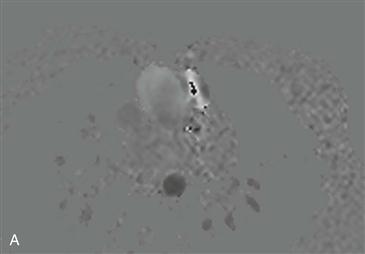
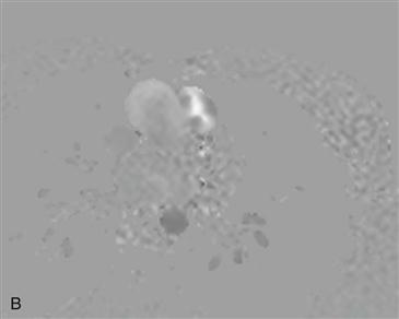
Two images from velocity-encoded cine phase contrast MRI demonstrate the imaging artifact of aliasing (Figs. A and B). Aliasing occurs when the encoded velocity set by the user is not higher than the actual velocity across the region of interest. These two images were acquired for quantification of known aortic stenosis. Cranial flow is depicted as white signal, and caudal flow is depicted as black signal. In the first image, the velocity-encoded gradient was set at 300 cm/s. There is black signal surrounded by white signal within the ascending aorta (Fig. A). This is physically impossible, and the imaging appearance is an aliasing artifact (wraparound or apparent velocity reversal). In the second image, the velocity-encoded gradient was increased to 350 cm/s, and the aliasing disappeared (Fig. B). The maximum velocity-encoded gradient should ideally be selected to exceed slightly the expected peak velocity.


