CASE 148
1. What should be included in the differential diagnosis? (Choose all that apply.)
C. Partial anomalous pulmonary venous connection (PAPVC)
2. Which septal defect is most commonly associated with PAPVC?
A. Secundum ASD
B. Primum ASD
3. What type of septal defect is seen in this patient?
A. Secundum ASD
B. Primum ASD
4. Which of the following most commonly occurs in the setting of long-standing ASD?
ANSWERS
CASE 148
Reference
Higgins CB. Radiography of congenital heart disease. In: Webb WR, Higgins CB, eds. Thoracic imaging: pulmonary and cardiovascular radiology. ed 2 Philadelphia: Lippincott Williams & Wilkins; 2010.
Cross-Reference
Cardiac Imaging: The REQUISITES, ed 3, pp 335–338.
Comment
Types of ASD
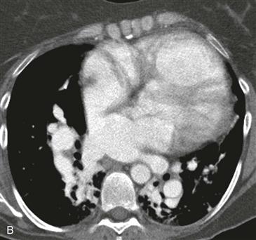
Types of ASD include ostium secundum, ostium primum, and sinus venosus. Ostium secundum ASD is the most common type and is the most frequently diagnosed left-to-right shunt in adult patients. An ostium primum ASD is present in atrioventricular septal defect (formerly known as endocardial cushion defect). A sinus venosus defect (Fig. B) occurs at the lateral superior portion of the ventricular septum, at the posterior wall of the superior vena cava, and is commonly associated with PAPVC. Patients with both PAPVC and ASD can be asymptomatic but may present with dyspnea, exercise intolerance, pulmonary hypertension, or syncope depending on the degree of shunt.
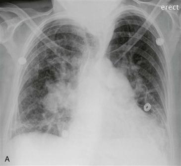
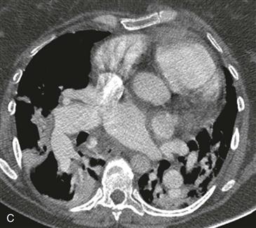
Imaging Findings
Although the chest radiograph may be normal when the shunt is small, pulmonary vascularity is usually increased (shunt vascularity) (Fig. A). PAPVC is another atrial-level shunt that can mimic an ASD physiologically and can appear similar to ASD on a chest radiograph. Echocardiography can delineate the size and location of the ASD. MRI or CT can be performed if echocardiography does not demonstrate a suspected ASD (Fig. B) and can be used to identify anomalous pulmonary veins (Fig. C). MRI can quantify the shunt fraction and helps determine the ideal timing to perform surgery. The indications for surgical correction are symptoms or a large shunt (i.e., pulmonary blood flow twice that of systemic blood flow).


